. 2014 Jul;92(7):605-12.
doi: 10.1139/cjpp-2014-0017. Epub 2014 Jun 18.

Impairment of autophagy in endothelial cells prevents shear-stress-induced increases in nitric oxide bioavailability

Affiliations

Impairment of autophagy in endothelial cells prevents shear-stress-induced increases in nitric oxide bioavailability

Leena P Bharath et al. Can J Physiol Pharmacol. 2014 Jul.

Abstract

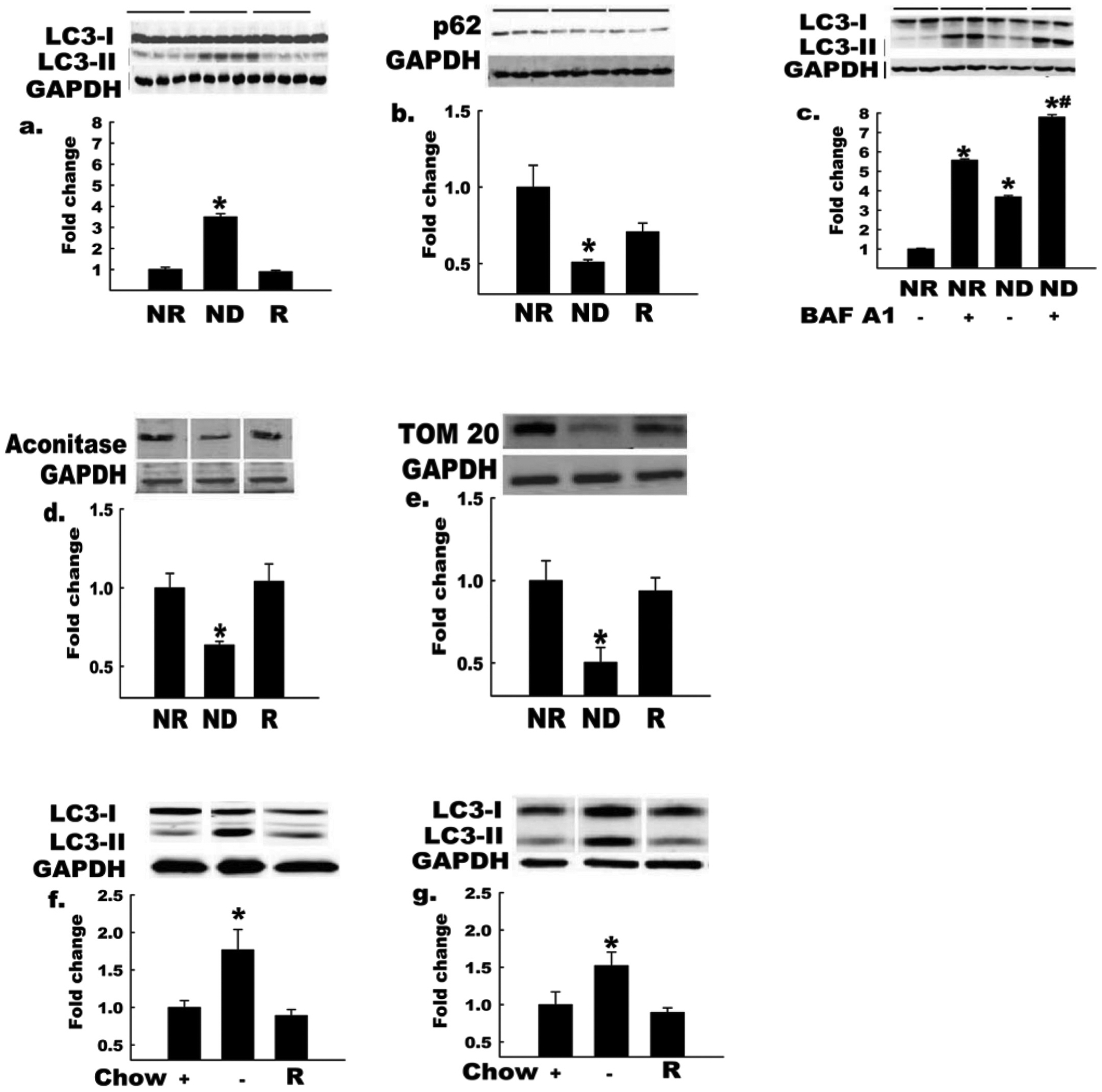
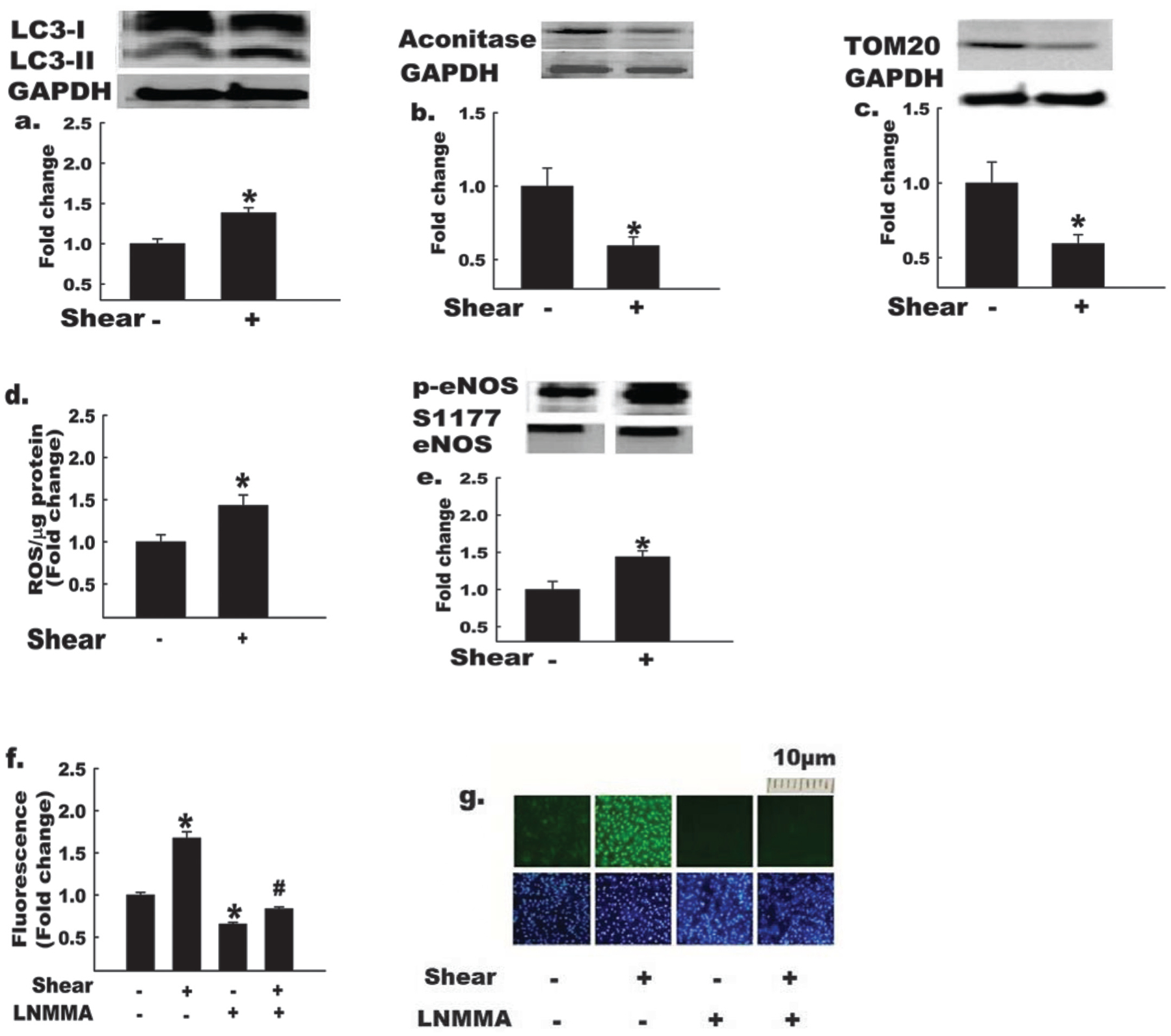
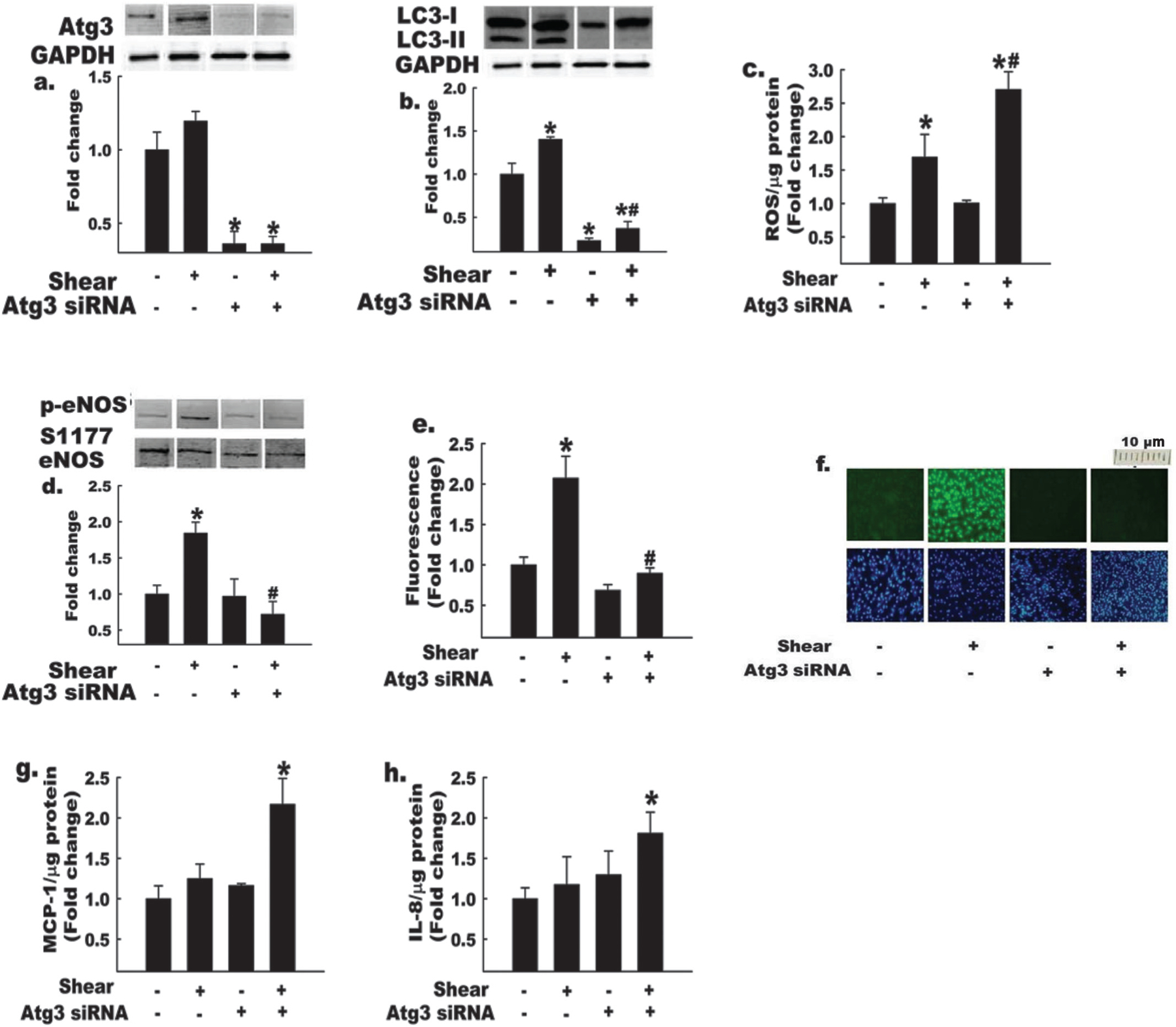
Autophagy is a lysosomal catabolic process by which cells degrade or recycle their contents to maintain cellular homeostasis, adapt to stress, and respond to disease. Impairment of autophagy in endothelial cells studied under static conditions results in oxidant stress and impaired nitric oxide (NO) bioavailability. We tested the hypothesis that vascular autophagy is also important for induction of NO production caused by exposure of endothelial cells to shear stress (i.e., 3 h × ≈20 dyn/cm(2)). Atg3 is a requisite autophagy pathway mediator. Control cells treated with non-targeting control siRNA showed increased autophagy, reactive oxygen species (ROS) production, endothelial NO synthase (eNOS) phosphorylation, and NO production upon exposure to shear stress (p < 0.05 for all). In contrast, cells with >85% knockdown of Atg3 protein expression (via Atg3 siRNA) exhibited a profound impairment of eNOS phosphorylation, and were incapable of increasing NO in response to shear stress. Moreover, ROS accumulation and inflammatory cytokine production (MCP-1 and IL-8) were exaggerated (all p < 0.05) in response to shear stress. These findings reveal that autophagy not only plays a critical role in maintaining NO bioavailability, but may also be a key regulator of oxidant-antioxidant balance and inflammatory-anti-inflammatory balance that ultimately regulate endothelial cell responses to shear stress.

L’autophagie est un processus catabolique lysosomal par lequel les cellules dégradent ou recyclent leur contenu afin de maintenir l’homéostasie cellulaire, s’adapter au stress et répondre à une maladie. La défaillance de l’autophagie chez les cellules endothéliales étudiées en conditions statiques provoque un stress oxydant et une diminution de la biodisponibilité d’oxyde nitrique (NO). Les auteurs ont testé l’hypothèse que l’autophagie vasculaire est aussi importante à l’induction de la production de NO provoquée par l’exposition des cellules endothéliales à un stress de cisaillement (par example, 3 h × ≈ 20 dyn/cm2). Atg3 est un médiateur nécessaire de la voie de l’autophagie. L’autophagie, la production d’espèces réactives d’oxygène (ERO), la phosphorylation de la NO synthase endothéliale (eNOS) et la production de NO étaient accrues chez des cellules contrôles traitées avec un pARNi contrôle non-ciblant, exposées à un stress de cisaillement (p < 0,05 pour tous les paramètres). En revanche, la phosphorylation de eNOS était diminuée de façon marquée chez les cellules dont Atg3 avait été inactivé de > 85 % par « knockdown » (au moyen du pARNi Atg3), et ces cellules étaient incapables d’accroitre le niveau de NO en réponse à un stress de cisaillement. De plus, l’accumulation d’ERO et la production de cytokines inflammatoires (MCP-1 et IL-8) étaient accrues (p < 0,05) en réponse à un stress de cisaillement. Ces données révèlent que l’autophagie joue non seulement un rôle dans le maintien de la biodisponibilité de NO, mais qu’elle agit aussi comme régulateur clé de la balance oxydante–anti-oxydante et inflammatoire–anti-inflammatoire, qui régule ultimement les réponses des cellules endothéliales au stress de cisaillement. [Traduit par la Rédaction]

Keywords: blood flow; exercice; exercise; flux sanguin; mitochondrial turnover; mitophagie; mitophagy; nutrient deprivation; oxidant stress; privation nutritionnelle; stress oxydant; vasculaire; vascular; « turnover » mitochondrial.


Figures

Fig. 1.
Fig. 1.
Autophagy is increased in endothelial cells and intact arteries by nutrient deprivation. Bovine aortic endothelial cells (BAECs) incubated in nutrient-deplete (ND) or nutrient-replete (NR) medium for 2 h. GAPDH, glyceraldehyde-3-phosphate dehydrogenase. LC3-II:LC3-I ratio (a) increased and p62 protein level (b) decreased (*, p < 0.05) in BAECs incubated in ND vs. NR medium (n = 4). These responses were restored when nutrients were added back to ND medium (R, third histogram). Horizontal bars above the images indicate the respective groups. LC3-II:LC3-I ratio (c) showed a greater increase in BAECs incubated with ND medium in the presence of the lysosomal proton pump inhibitor BAF A1, owing to the expected effect of this treatment to block lysosomal degradation of LC3-II (n = 3 per condition; *, p < 0.05 for BAF A1 effect; #, p < 0.05 for −AA effect). Protein levels of the mitochondrial markers m-aconitase and TOM 20 (d, e) decreased in cells incubated in ND vs. NR medium (n = 4, p < 0.05), suggesting increased mitochondrial turnover; in both cases (d, e), responses were reversed when nutrients were restored to ND medium (R). LC3-II:LC3-I increased (*, p < 0.05) in hearts (f) and arteries (g) from in vivo fasted (14 h) vs. fed mice, and these responses were reversed by 1 h re-feeding (R, n = 3 per group). Histograms (below) represent the mean ± SE of densitometry (above). *, p < 0.05 for ND medium (d,e) or fasting (Chow −) (f, g) effect. For Figs. 1a–1e, each n refers to one 10 cm Petri dish. For Figs. 1f and 1g, each n refers to one mouse.
Fig. 2.
Fig. 2.
Autophagy and nitric oxide (NO) bioavailability are elevated in endothelial cells exposed to shear stress. Bovine aortic endothelial cells (BAECs) were exposed to shear stress (shear +) or static conditions (shear −) for 3 h, as indicated. GAPDH, glyceraldehyde-3-phosphate dehydrogenase. Shear + BAECs show increased (*, p < 0.05) LC3-II:LC3-I ratio (a), and decreased protein levels of mitochondrial markers, m-aconitase (b), and TOM20 (c); in addition, shear + BAECs exhibit increased reactive oxygen species (ROS) production (d), p-eNOS S1177 : total eNOS (e), and NO production (mean data (panel f), representative images of DAF-FM fluorescence (top row, panel g)) in comparison with shear – BAECs (n = 4–6 per experiment). Treatment of BAECs with the NO synthase inhibitor, monomethyl-L-arginine (L-NMMA), reduced (p < 0.05) NO production ± shear stress (mean data (panel f), representative images of DAF-FM (panel g, top row) and DAPI nuclear fluorescence (panel g, bottom row)). The bottom row of panel g indicates that cell density was similar among treatments. For Figs. 2a, 2b, 2c, and 2e, the histograms (below) represent the mean ± SE of densitometry (above). *, p < 0.05 for shear effect; #, p < 0.05 for L-NMMA effect. For Figs. 2a, 2b, 2c, and 2e, each n refers to one 10 cm petri dish. For Figs. 2d, 2f, and 2g, each n refers to one well of a 6-well plate.
Fig. 3.
Fig. 3.
Impairment of autophagy impairs shear-stress-induced increases in eNOS S1177 phosphorylation and nitric oxide (NO) bioavailability. Bovine aortic endothelial cells (BAECs) treated with Atg3 siRNA (+) or control scrambled siRNA (−) were exposed to shear stress (shear +) or static conditions (shear −), as indicated. Atg3 protein knockdown efficiency (a) was identical for each condition (*, p < 0.05 for Atg3 siRNA effect; p = NS for static vs. shear-stress comparisons). Basal and shear-stress-induced autophagy (assessed by LC3-II : LC3-I ratio) were suppressed + Atg3 siRNA (b) under both static and shear-stress conditions. In addition, Atg3 siRNA-treated BAECs exhibited increased (c) reactive oxygen species (ROS) production and decreased (d) p-eNOS S1177 : total eNOS in response to shear stress. NO levels (e) were decreased in Atg3 siRNA-treated BAECs ± shear stress. Mean data (panel e) and representative images of DAF-FM (panel f, top row) and DAPI nuclear fluorescence (panel f, bottom row) are shown. The bottom row of panel f indicates that cell density was similar among treatments. Atg3 siRNA-treated BAECs exhibit increased expression of proinflammatory cytokines (g) MCP-1 and (h) IL8, after exposure to shear stress. For Figs. 3a, 3b, and 3d, histograms (below) represent the mean ± SE of densitometry of 4–6 experiments (above). *, p < 0.05 for shear effect; #, p < 0.05 for Atg3 effect. For Figs. 3a, 3b, and 3d, each n refers to one 10 cm petri dish. For Figs. 3c, 3e, 3f, 3g, and 3h, each n refers to one well of a 6-well plate.

References

    1. Babu PV, Si H, Fu Z, Zhen W, and Liu D 2012a. Genistein prevents hyperglycemia-induced monocyte adhesion to human aortic endothelial cells through preservation of the cAMP signaling pathway and ameliorates vascular inflammation in obese diabetic mice. J. Nutr 142(4): 724–730. doi: 10.3945/jn.111.152322. - DOI - PMC - PubMed
    1. Babu PV, Si H, and Liu D 2012b. Epigallocatechin gallate reduces vascular inflammation in db/db mice possibly through an NF-kappaB-mediated mechanism. Mol. Nutr. Food Res 56(9): 1424–1432. doi:10.1002/mnfr.201200040. - DOI - PMC - PubMed
    1. Balaban RS, Nemoto S, and Finkel T 2005. Mitochondria, oxidants, and aging. Cell, 120(4): 483–495. doi:10.1016/j.cell.2005.02.001. - DOI - PubMed
    1. Bensaad K, Cheung EC, and Vousden KH 2009. Modulation of intracellular ROS levels by TIGAR controls autophagy. EMBO J. 28(19): 3015–3026. doi:10.1038/emboj.2009.242. - DOI - PMC - PubMed
    1. Bevan HS, Slater SC, Clarke H, Cahill PA, Mathieson PW, Welsh GI, et al.2011. Acute laminar shear stress reversibly increases human glomerular endothelial cell permeability via activation of endothelial nitric oxide synthase. Am. J. Physiol. Renal Physiol 301(4): F733–F742. doi:10.1152/ajprenal.00458.2010. - DOI - PMC - PubMed

Publication types

LinkOut - more resources