. 2014 Oct;76(10):1329-37.
doi: 10.1292/jvms.14-0260. Epub 2014 Jun 19.

Effects of exposure to male goat hair extracts on luteinizing hormone secretion and neuronal activation in seasonally anestrous ewes

Affiliations

Effects of exposure to male goat hair extracts on luteinizing hormone secretion and neuronal activation in seasonally anestrous ewes

Hiromi Ohara et al. J Vet Med Sci. 2014 Oct.

Abstract

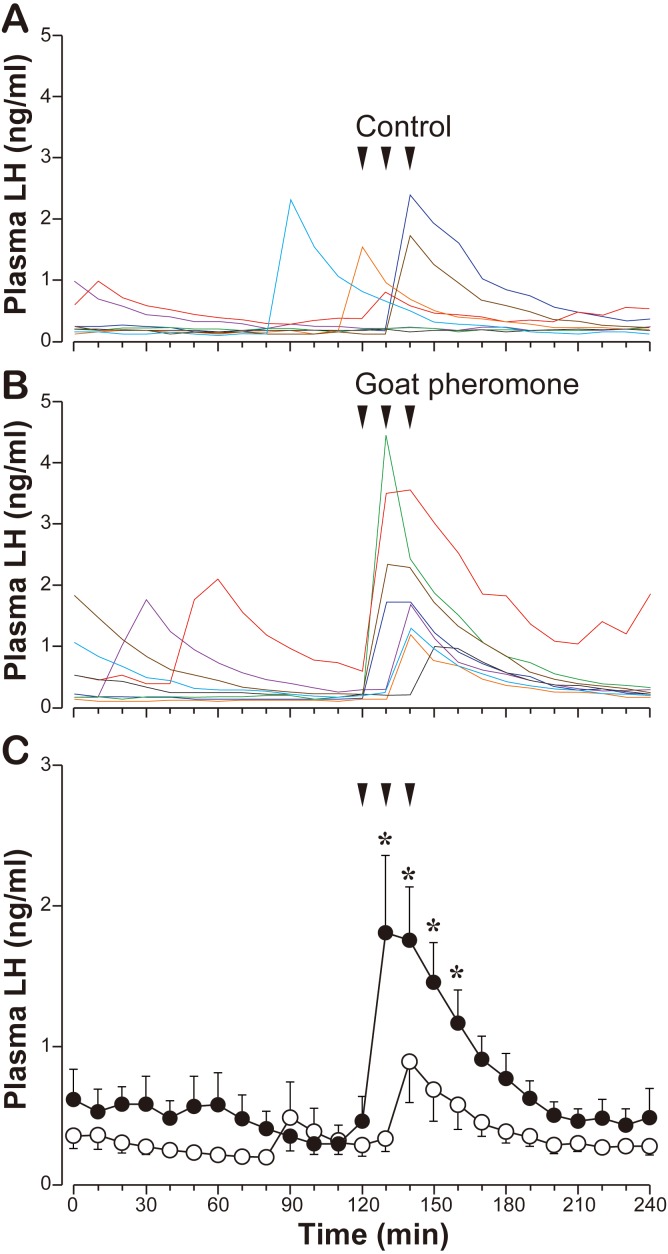
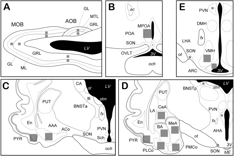
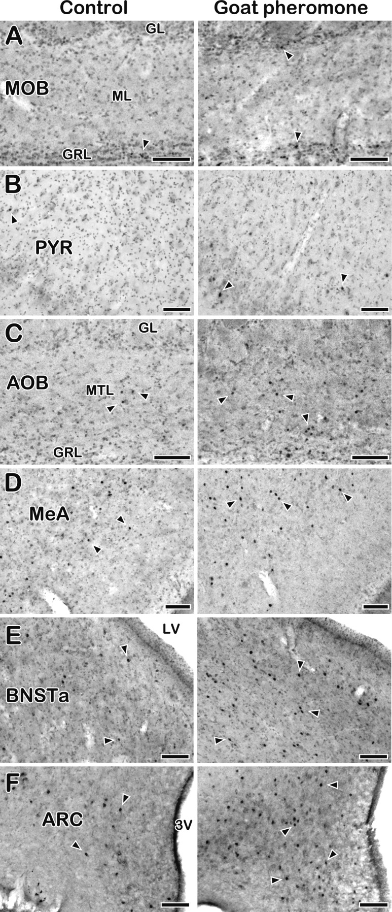
In sheep and goats, exposure of seasonally anestrous females to males or their fleece/hair activates the gonadotropin-releasing hormone (GnRH) pulse generator leading to pulsatile luteinizing hormone (LH) secretion. Pheromones emitted by sexually mature males are thought to play a prominent role in this male effect. In the present study, we first aimed to clarify whether the male goat pheromone is effective in ewes. Seasonally anestrous St. Croix ewes were exposed to hair extracts derived from either intact or castrated (control) male Shiba goats. The male goat-hair extract significantly increased LH secretion compared to the control, suggesting that an interspecies action of the male pheromone occurs between sheep and goats. Using the male goat-hair extract as the pheromone source, we then aimed to clarify the neural pathway involved in the signal transduction of the male pheromone. Ewes were exposed to either the goat-hair extract or the control and sacrificed 2 hr after the exposure. Expression of c-Fos, a marker of neuronal activation, was immunohistochemically examined. The male goat-hair extract significantly increased the c-Fos expression compared to the control in regions of the vomeronasal system, such as the accessory olfactory bulb and medial amygdala, and the arcuate nucleus. The main olfactory bulb did not exhibit any significant increase in the c-Fos expression by the male goat-hair extract. This result suggests that the neural signal of the male pheromone is conveyed to the GnRH pulse generator through the activated regions in ewes.


Figures

Fig. 1.
Fig. 1.
Effects of the goat pheromone exposure on LH secretion in seasonally anestrous ewes. A, individual profiles (n=8) of LH concentrations in the control (the hair extract of castrated male goats) exposure. B, individual profiles (n=8) of LH concentrations in the goat pheromone (the hair extract of intact male goats) exposure. Lines with the same color in A and B indicate the results of the same individual. C, the mean ± SEM of LH concentrations in each treatment. Open circles, the control exposure. Solid circles, the goat pheromone exposure. Arrows indicate timing of exposure. *, significantly different (P<0.05, Student’s t-tests) compared to the corresponding control value.
Fig. 2.
Fig. 2.
Schematic illustrations of brain regions. A, a sagittal view of the olfactory bulb. B–E, coronal views of the hypothalamus and the amygdaloid complex (the rostro-caudal order). Gray squares schematically show the areas in which the number of c-Fos positive cells was counted in each brain region. AAA, anterior amygdaloid area; ACo, anterior cortical amygdala; AHA, anterior hypothalamic area; AOB, accessory olfactory bulb; ARC, arcuate nucleus; BA, basal amygdala, BNSTa, anterior part of the bed nucleus of the stria terminalis; BNSTp, posterior part of the bed nucleus of the stria terminalis; CA, caudate nucleus; CeA, central amygdala; DMH, dorsomedial hypothalamic nucleus; En, endopiriform nucleus; LA, lateral amygdala; LHA, lateral hypothalamic area; MeA, medial amygdala; MOB, main olfactory bulb; MPOA, medial preoptic area; OVLT, vascular organ of the lamina terminalis; PMCo, posterior medial cortical amygdala; PLCo, posterior lateral cortical amygdala; POA, preoptic area; PUT, putamen; PVN, paraventricular nucleus of the hypothalamus; PYR, piriform cortex; Sch, suprachiasmatic nucleus; SON, supraoptic nucleus; VMH, ventromedial hypothalamic nucleus, GL, glomerular layer; GRL, granule cell layer; ML, mitral cell layer; MTL, mitral/tufted cell layer; ac, anterior commissure; fx, fornix; och, optic chiasm; ot, optic tract; st, stria terminalis; stm, stria medullaris of the thalamus; LV, lateral ventricle; ME, median eminence; 3V, third ventricle.
Fig. 3.
Fig. 3.
Photomicrographs of c-Fos-ir cells in several brain regions in seasonally anestrous ewes exposed to the control (left column) or goat pheromone (right column). A, main olfactory bulb (MOB). B, piriform cortex (PYR). C, accessory olfactory bulb (AOB). D, medial amygdala (MeA). E, anterior part of the bed nucleus of the stria terminalis (BNSTa). F, arcuate nucleus (ARC). Sections were briefly counter-stained for Nissl. Arrowheads indicate representative c-Fos-ir materials. GL, glomerular layer; GRL, granule cell layer; ML, mitral cell layer; MTL, mitral/tufted cell layer, LV, lateral ventricle; 3V, third ventricle. Scale bars, 100 µm.

References

    1. Blache D., Fabre-Nys C. J., Venier G.1991. Ventromedial hypothalamus as a target for oestradiol action on proceptivity, receptivity and luteinizing hormone surge of the ewe. Brain Res. 546: 241–249. doi: 10.1016/0006-8993(91)91488-M - DOI - PubMed
    1. Brennan P. A.2001. The vomeronasal system. Cell. Mol. Life Sci. 58: 546–555. doi: 10.1007/PL00000880 - DOI - PMC - PubMed
    1. Bressler S. C., Baum M. J.1996. Sex comparison of neuronal Fos immunoreactivity in the rat vomeronasal projection circuit after chemosensory stimulation. Neuroscience 71: 1063–1072. doi: 10.1016/0306-4522(95)00493-9 - DOI - PubMed
    1. Chemineau P.1987. Possibilities for using bucks to stimulate ovarian and oestrous cycles in anovulatory goats— a review. Livest. Prod. Sci. 17: 135–147. doi: 10.1016/0301-6226(87)90059-5 - DOI
    1. Clark A. S., Pfeifle J. K., Edwards D. A.1981. Ventromedial hypothalamic damage and sexual proceptivity in female rats. Physiol. Behav. 27: 597–602. doi: 10.1016/0031-9384(81)90228-6 - DOI - PubMed

Publication types

Substances