

Towards highly accelerated Cartesian time-resolved 3D flow cardiovascular magnetic resonance in the clinical setting
- PMID: 24942253
- PMCID: PMC4230248
- DOI: 10.1186/1532-429X-16-42
Towards highly accelerated Cartesian time-resolved 3D flow cardiovascular magnetic resonance in the clinical setting
Abstract
Background: The clinical applicability of time-resolved 3D flow cardiovascular magnetic resonance (CMR) remains compromised by the long scan times associated with phase-contrast imaging. The present work demonstrates the applicability of 8-fold acceleration of Cartesian time-resolved 3D flow CMR in 10 volunteers and in 9 patients with different congenital heart diseases (CHD). It is demonstrated that accelerated 3D flow CMR data acquisition and image reconstruction using k-t PCA (principal component analysis) can be implemented into clinical workflow and results are sufficiently accurate relative to conventional 2D flow CMR to permit for comprehensive flow quantification in CHD patients.
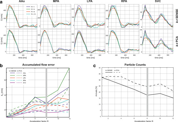
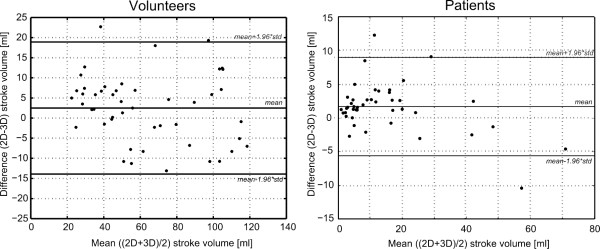
Methods: The fidelity of k-t PCA was first investigated on retrospectively undersampled data for different acceleration factors and compared to k-t SENSE and fully sampled reference data. Subsequently, k-t PCA with 8-fold nominal undersampling was applied on 10 healthy volunteers and 9 CHD patients on a clinical 1.5 T MR scanner. Quantitative flow validation was performed in vessels of interest on the 3D flow datasets and compared to 2D through-plane flow acquisitions. Particle trace analysis was used to qualitatively visualise flow patterns in patients.
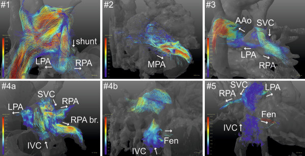
Results: Accelerated time-resolved 3D flow data were successfully acquired in all subjects with 8-fold nominal scan acceleration. Nominal scan times excluding navigator efficiency were on the order of 6 min and 7 min in patients and volunteers. Mean differences in stroke volume in selected vessels of interest were 2.5 ± 8.4 ml and 1.63 ± 4.8 ml in volunteers and patients, respectively. Qualitative flow pattern analysis in the time-resolved 3D dataset revealed valuable insights into hemodynamics including circular and helical patterns as well as flow distributions and origin in the Fontan circulation.
Conclusion: Highly accelerated time-resolved 3D flow using k-t PCA is readily applicable in clinical routine protocols of CHD patients. Nominal scan times of 6 min are well tolerated and allow for quantitative and qualitative flow assessment in all great vessels.
Figures
References
-
- Nordmeyer S, Riesenkampff E, Crelier G, Khasheei A, Schnackenburg B, Berger F, Kuehne T. Flow‐sensitive four‐dimensional cine magnetic resonance imaging for offline blood flow quantification in multiple vessels: a validation study. J. Magn. Reson. Imaging. 2010;32:677–683. - PubMed
-
- Markl M, Wallis W, Brendecke S, Simon J, Frydrychowicz A, Harloff A. Estimation of global aortic pulse wave velocity by flow‐sensitive 4D MRI. Magn. 2010;63:1575–1582. - PubMed
-
- Ebbers T, Wigström L, Bolger AF, Engvall J, Karlsson M. Estimation of relative cardiovascular pressures using time‐resolved three‐dimensional phase contrast MRI. Magn. 2001;45:872–879. - PubMed
-
- Stalder A, Russe M, Frydrychowicz A, Bock J, Hennig J, Markl M. Quantitative 2D and 3D phase contrast MRI: optimized analysis of blood flow and vessel wall parameters. Magn. 2008;60:1218–1231. - PubMed
Publication types
MeSH terms
Grants and funding
LinkOut - more resources
Full Text Sources
Other Literature Sources
Medical
