. 2014 Sep 1;88(17):9830-41.
doi: 10.1128/JVI.01425-14. Epub 2014 Jun 18.

Enterovirus 71 3C inhibits cytokine expression through cleavage of the TAK1/TAB1/TAB2/TAB3 complex

Affiliations

Enterovirus 71 3C inhibits cytokine expression through cleavage of the TAK1/TAB1/TAB2/TAB3 complex

Xiaobo Lei et al. J Virol. .

Abstract

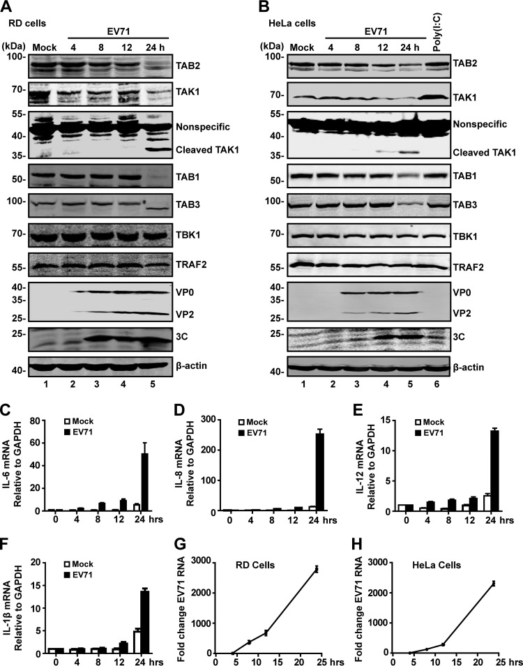
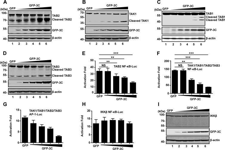
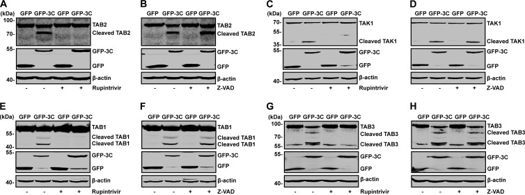
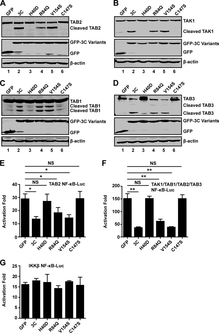
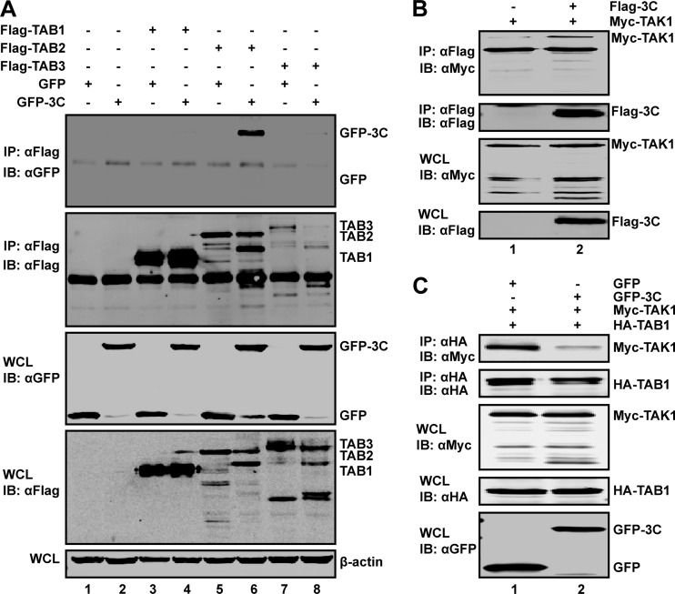
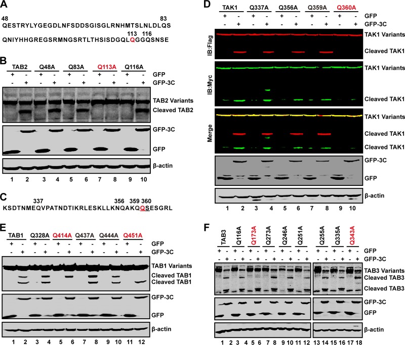
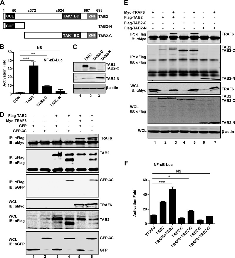
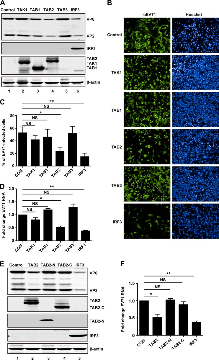
Enterovirus 71 (EV71) causes hand, foot, and mouth disease in young children and infants. Severe infection with EV71 can lead to various neurological complications or fatal diseases. However, the mechanism of EV71 pathogenesis is poorly understood. Emerging evidence suggests that EV71 modulates type I interferon (IFN) and cytokine responses. Here, we show that EV71 disables components of the TAB2 complex through the 3C protein. When expressed in mammalian cells, EV71 3C interacts with TAB2 and TAK1, which inhibits NF-κB activation. Furthermore, 3C mediates cleavage of TAB2 and its partners, which requires the protease activity. H40D or C147S substitution in the 3C active sites abolishes its activity, whereas R84Q or V154S substitution in the RNA binding domain has no effect. The 3C protein targets TAB2 at Q113-S114, TAK1 at Q360-S361, TAB1 both at Q414-G415 and Q451-S452, and TAB3 at Q173-G174 and Q343-G344. Importantly, overexpression of TAB2 inhibits EV71 replication, whereas addition of cleaved fragments has no effect. Thus, an equilibrium between the TAB2 complex and EV71 3C represents a control point of viral infection. These results suggest that TAK1/TAB1/TAB2/TAB3 cleavage mediated by EV71 may be a mechanism to interfere with inflammatory responses.

Importance: The TAK1 complex plays a critical role in the activation of NF-κB and cytokine production. However, little is known about its connection to enterovirus 71 (EV71). We demonstrate that EV71 3C suppresses cytokine expression via cleavage of the TAK1 complex proteins. EV71 3C interacts with TAB2 and TAK1. Furthermore, overexpression of TAB2 inhibits EV71 replication, whereas addition of cleaved fragment has no effect. These results suggest that the interplay of EV71 and the TAK1 complex influences the outcome of viral infection.


Figures

FIG 1
FIG 1
EV71 infection induces cleavage of TAB2, TAK1, TAB1, and TAB3. RD (A) or HeLa (B) cells were mock infected or infected with EV71 (MOI of 1 PFU per cell for RD; MOI of 2 PFU per cell for HeLa). HeLa cells were treated with poly(I·C) for 4 h, which was added to the medium. At 4, 8, 12, and 24 h postinfection, cell lysates were prepared and analyzed by Western blotting with antibodies for TAB2, TAK1, TAB1, TAB3, TBK1, TRAF2, EV71, EV71-3C, and β-actin. Data are representative of three independent experiments. (C to F) HeLa cells were mock infected or infected with EV71. At 24 h after infection, total RNA extracted for cells was analyzed for the expression of IL-6 (C), IL-8 (D), IL-12 (E), and IL-1β (F) by quantitative real-time PCR using SYBR green. (G and H) RD cells or HeLa cells were infected with EV71. At different time points after infection, total RNA was extracted, and the viral RNA levels of EV71 were evaluated by quantitative real-time PCR using SYBR green. Data are expressed as fold change of the EV71 RNA level relative to that at the 4-h time point using the ΔΔCT method as described in Materials and Methods.
FIG 2
FIG 2
The 3C protease of EV71 mediates cleavage of TAK1 complex components. (A to D) 293T cells were transfected with plasmids encoding TAB2 (A), TAK1 (B), TAB1(C), or TAB3 (D), along with GFP (lane 1) or increasing amounts of GFP-3C (lanes 2 to 6). At 24 h after transfection, lysates of cells were analyzed by Western blotting with antibodies against Flag, GFP, Myc, or β-actin. β-Actin was used as a protein loading control. (E and F) EV71 3C inhibits NF-κB promoter activation. 293T cells were transfected with TAB2 alone or a mixture of TAK1, TAB1, TAB2, and TAB3, along with NF-κB-Luc and increasing amounts of GFP-3C. A plasmid expressing GFP or pRL-SV40 was used as a control. At 24 h after transfection, cell lysates were assayed for luciferase activities. (G) EV71 3C inhibits AP-1 promoter activation. 293T cells were transfected with TAK1, TAB1, TAB2 and TAB3, along with AP-1-Luc and increasing amounts of GFP-3C. Data are a representative of three independent experiments with triplicate samples. (H) 293T cells were transfected with plasmids encoding IKKβ, along with NF-κB-Luc and increasing amounts of GFP-3C plasmid. At 24 h after transfection, cell lysates were assayed for luciferase activities. (I) Cell lysates from panel H were analyzed by Western blotting with antibodies against Flag, GFP, or β-actin, respectively. **, P < 0.01; ***, P < 0.001; NS, nonsignificant.
FIG 3
FIG 3
Effects of selected inhibitors on cleavage of TAB2, TAK1, TAB1, and TAB3. 293T cells were transfected with plasmids encoding TAB2 (A and B), TAK1 (C and D), TAB1 (E and F), and TAB3 (G and H), along with plasmids encoding GFP or GFP-3C. At 4 h after transfection, cells were incubated with the protease inhibitor rupintrivir (A, C, E, and G) (2 nM) or caspase inhibitor Z-VAD (B, D, F, and H) (20 μM). At 24 h after transfection, cell lysates were then processed for Western blot analysis with antibodies specific for Flag, GFP, or Myc.
FIG 4
FIG 4
Mutational effects on EV71 3C activities. 293T cells were transfected with plasmids encoding TAB2(A), TAK1 (B), TAB1 (C), or TAB3 (D), along with GFP (lane 1) or GFP-3C variants (lanes 2 to 6), as indicated. At 24 h after transfection, cell lysates were analyzed by Western blotting using a Li-Cor Odyssey Dual-Color System (Li-Cor, Lincoln, NE). (E, F, and G) 293T cells were transfected with the TAB2 (E), TAK1 complex (F), or IKKβ (G), along with NF-κB-Luc and GFP-3C variants. A plasmid expressing GFP or pRL-SV40 was used as a control. At 24 h after transfection, cell lysates were assayed for luciferase activities. Data are representative of three independent experiments with triplicate samples. *, P < 0.05; **, P < 0.01; NS, nonsignificant.
FIG 5
FIG 5
(A) EV71 3C associates with TAB2. 293T cells were transfected with plasmids encoding TAB1 (lanes 3 and 4), TAB2 (lanes 5 and 6), TAB3 (lanes 7 and 8), GFP (lanes 1, 3, 5, and 7), and GFP-3C (lanes 2, 4, 6, and 8). Empty pCMV6 (lanes 1 and 2) vector was used as a control. At 24 h after transfection, cell lysates were immunoprecipitated with antibody against Flag. Samples were then subjected to Western blot analysis. (B) EV71 3C interacts with TAK1. 293T cells were transfected with plasmids encoding Myc-TAK1 (lanes 1 and 2) and Flag-3C (lane 2). The total amount of DNA was kept constant using empty pCDNA3.1 (lane 1). At 24 h after transfection, cell lysates were immunoprecipitated with Flag antibody. Immunoprecipitates and cell lysates were then subjected to Western blot analysis. (C) EV71 3C inhibits the interaction between TAK1 and TAB1. 293T cells were transfected with plasmids encoding Myc-TAK1 (lanes 1 and 2), hemagglutinin (HA)-TAB1 (lanes 1 and 2), and GFP-3C (lane 2). At 24 h after transfection, cell lysates were immunoprecipitated with HA antibody. Immunoprecipitates and cell lysates were then subjected to Western blot analysis. IB, immunoblotting; IP, immunoprecipitation; WCL, whole-cell lysate; α, anti.
FIG 6
FIG 6
The cleavage sites of 3C in TAK1 complex. (A) Primary sequences of amino acids 48 to 120 within TAB2. In this region, glutamines of 48, 83, 113, and 116 sites were replaced with alanines. (B) The 3C cleavage site in TAB2. 293T cells were transfected with plasmids encoding wild-type TAB2 or TAB2 variants as indicated along with GFP (lanes 1, 3, 5, 7, and 9) or GFP-3C (lanes 2, 4, 6, 8, and 10). At 24 h after transfection, cell lysates were subjected to Western blot analysis using antibodies against Flag and GFP. β-Actin was used as a loading control. (C) Primary sequence of amino acids 330 to 366 within TAK1. In this region, glutamines were replaced with alanines. (D) The 3C cleavage site in TAK1. 293T cells were transfected with wild-type TAK1 or TAK1 mutants as indicated along with GFP (lanes 1, 3, 5, 7, and 9) or GFP-3C (lanes 2, 4, 6, 8, and 10). At 24 h after transfection, cell lysates were subjected to Western blot analysis with antibodies using a Li-Cor Odyssey Dual-Color System. Two antibodies that recognize TAK1 (Myc, C terminus of TAK1, 800 nm, green; Flag, N terminus of TAK1, 700 nm, red) were used. β-Actin was used as a loading control. (E and F) The 3C cleavage sites in TAB1 and TAB3. 293T were transfected with wild-type TAB1 or its variants (E) or TAB3 or its variants (F) as indicated along with GFP or GFP-3C. Cell lysates were subjected to Western blot analysis.
FIG 7
FIG 7
(A) Schematic diagrams of TAB2 and deletion mutants. Domain organization is outlined: the binding ubiquitin-conjugating enzyme domain (CUE), the zinc finger domain (ZNF), and phosphoserine (S372 and S524). TAB2-N and TAB2-C represent cleaved fragments of TAB2 by 3C. (B) The effect of TAB2 and its deletion mutants on NF-κB promoter activation. 293T cells were transfected with plasmids encoding TAB2, TAB2-N, or TAB2-C, along with NF-κB-Luc. pRL-SV40 was used as a control. At 24 h after transfection, cell lysates were assayed for luciferase activities. Data are representative of three independent experiments with triplicate samples. (C) Expression of TAB2 mutants. Cell lysates from panel B were subjected to Western blot analysis with antibodies against Flag and β-actin. (D) 293T cells were transfected with plasmids encoding TAB2 (lanes 3, 4, 5, and 6), TRAF6 (lanes 5 and 6), GFP (lanes 1, 3, and 5), and GFP-3C (lanes 2, 4, and 6). At 24 h after transfection, cell lysates were immunoprecipitated with antibody against Flag. Samples were then subjected to Western blot analysis. (E) 293T cells were transfected with plasmids encoding TRAF6 (lanes 1, 3, 5, and 7), TAB2 (lanes 2 and 3), TAB2-C (lanes 4 and 5), and TAB2-N (lanes 6 and 7). At 24 h after transfection, cell lysates were immunoprecipitated and detected as described in panel D. (F) 293T cells were transfected with plasmids as indicated, along with NF-κB-Luc. pRL-SV40 was used as a control. At 24 h after transfection, cell lysates were assayed for luciferase activities. *, P < 0.05; **, P < 0.01; ***, P < 0.001; NS, nonsignificant.
FIG 8
FIG 8
(A) TAB2 inhibits EV71 replication. RD cells were transfected with plasmids encoding Flalg-TAK1, Flag-TAB1, Flag-TAB2, or Flag-TAB3. Myc-IRF3 used as a control. At 24 h after transfection, cells were treated with EV71 at an MOI of 1 PFU per cell. After 24 h cells were harvested and resolved with 12% SDS-PAGE. Western blot analysis for EV71, Flag, Myc, or β-actin was conducted. (B) RD cells were transfected with plasmids encoding TAK1, TAB1, TAB2, TAB3, or IRF3. At 24 h after transfection, cells were treated with EV71. After 24 h, cells were fixed and stained with antibody against EV71. The images were acquired on an Operetta instrument using a 20× long-working-distance objective. Representative images are shown. (C) The percentage of EV71-infected cells from the experiment shown in panel B was analyzed using Harmony software. (D) RD cells were transfected with plasmids encoding TAK1, TAB1, TAB2, TAB3, or IRF3. Empty vector was used as a control. At 24 h after transfection, cells were infected with EV71. After 24 h, total RNA was extracted, and the viral RNA levels of EV71 were evaluated by quantitative real-time PCR using SYBR green. Data are expressed as fold change of the EV71 RNA level relative to the control using the ΔΔCT method as described in Materials and Methods. (E) RD cells were transfected with TAB2 variants as indicated. IRF3 was used as a control. Cell lysates were detected as described in panel A. (F) RD cells were transfected as described in panel E. At 24 h after transfection, cells were treated with EV71. After 24 h, EV71 RNA levels were detected and expressed as described in panel D. *, P < 0.05; **, P < 0.01; NS, nonsignificant.

Similar articles

Cited by

References

    1. McMinn PC. 2002. An overview of the evolution of enterovirus 71 and its clinical and public health significance. FEMS Microbiol. Rev. 26:91–107. 10.1111/j.1574-6976.2002.tb00601.x - DOI - PubMed
    1. Li ML, Hsu TA, Chen TC, Chang SC, Lee JC, Chen CC, Stollar V, Shih SR. 2002. The 3C protease activity of enterovirus 71 induces human neural cell apoptosis. Virology 293:386–395. 10.1006/viro.2001.1310 - DOI - PubMed
    1. Shih SR, Chiang C, Chen T, Wu CN, Hsu J, Lee J, Hwang MJ, Li M, Chen G, Ho MS. 2004. Mutations at KFRDI and VGK domains of enterovirus 71 3C protease affect its RNA binding and proteolytic activities. J. Biomed. Sci. 11:239–248. 10.1007/BF02256567 - DOI - PubMed
    1. Sim AC, Luhur A, Tan TM, Chow VT, Poh CL. 2005. RNA interference against enterovirus 71 infection. Virology 341:72–79. 10.1016/j.virol.2005.06.047 - DOI - PubMed
    1. Hung HC, Wang HC, Shih SR, Teng IF, Tseng CP, Hsu JT. 2011. Synergistic inhibition of enterovirus 71 replication by interferon and rupintrivir. J. Infect. Dis. 203:1784–1790. 10.1093/infdis/jir174 - DOI - PubMed

Publication types

MeSH terms