Review
. 2014 Jul;260(1):76-85.
doi: 10.1111/imr.12189.

Intestinal IgA production and its role in host-microbe interaction

Affiliations
Review

Intestinal IgA production and its role in host-microbe interaction

Cindy Gutzeit et al. Immunol Rev. 2014 Jul.

Abstract

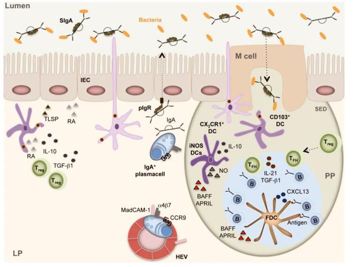
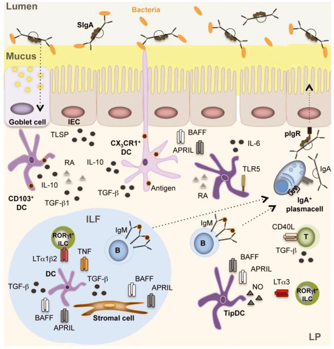
Complex and diverse communities of bacteria establish mutualistic and symbiotic relationships with the gut after birth. The intestinal immune system responds to bacterial colonization by acquiring a state of hypo-responsiveness against commensals and active readiness against pathogens. The resulting homeostatic balance involves a continuous dialog between the microbiota and lymphocytes with the intermediation of epithelial and dendritic cells. This dialog causes massive production of immunoglobulin A (IgA), a non-inflammatory antibody specialized in mucosal protection. Here, we discuss recent advances on the regulation of intestinal IgA responses and their role in host-microbe interaction.

Keywords: B cells; T cells; class switching; dendritic cells; epithelial cells; immunoglobulin; mucosal immunity.


Figures

Fig. 1
Fig. 1. Intestinal IgA production in Peyer’s patches (PPs)
M cells intercalated in the follicle-associated epithelium of PPs capture luminal SIgA-coated antigens through SIgA receptors. Luminal antigens are also captured by transepithelial projections emanating from non-migratory macrophage-like CX3CR1+ dendritic cells (DCs) in the subepithelial dome (SED) of PPs. Then, M cells and CX3CR1+ DCs transfer antigen to migratory CD103+ DCs, which move to the interfollicular area of PPs to establish cognate interactions with CD4+ T cells, including Treg cells. These tolerogenic CD4+ T cells have IgA-inducing function and require RA, TGF-β1, IL-10, and TSLP from intestinal epithelial cells (IECs) and DCs. Treg cells further differentiate to Tfh cells, which establish cognate interactions with antigen-specific B cells at the T-B cell border. Tfh cellactivated B cells migrate to the follicle together with Tfh cells by following a CXCL13 gradient formed by follicular dendritic cells (FDCs). In the presence of antigen-retaining FDCs, B cells undergo expansion, SHM, IgA CSR, and affinity maturation in response to CD40L, IL-21, and TGF-β1 from Tfh cells. TGF-β1, IL-10, RA, and nitric oxide (NO) from FDCs and TipDCs further enhance IgA CSR and production in the GC. B cells emerging from the GC upregulate α4β7 and CCR9 gut-homing receptors in response to RA, reach the general circulation, and finally enter the lamina propria (LP) through MAdCAM-1-expressing high endothelial venules. In the LP, B cells differentiate to plasmablasts and plasma cells that secrete polymeric IgA. Following pIgR-mediated transcytosis across IECs, polymeric IgA forms SIgA, which interacts with commensal bacteria and neutralizes pathogens.
Fig. 2
Fig. 2. Intestinal IgA production in isolated lymphoid follicles (ILFs) and lamina propria (LP)
CX3CR1+ dendritic cells (DCs) capture luminal antigens through transepithelial projections. Luminal antigens are also captured by CD103+ DCs through transient passages formed by MUC2-secreting goblet cells. These DCs receive conditioning TSLP, RA, TGF-β1, and MUC2 signals from intestinal epithelial cells (IECs) or goblet cells and then present antigen to B cells located in ILFs or in the diffuse lymphoid tissue of the LP. In ILFs, B cells receive IgA-inducing signals from BAFF, APRIL, and TGF-β1 produced by DCs and stromal cells in response to TNF and LT from LTi cells and TLR ligands from the microbiota. In the LP, B cells receive IgA-inducing signals from BAFF, APRIL, and nitric oxide (NO) produced by TipDCs and TLR ligands from the microbiota. Of note, NO enhances BAFF and APRIL produced by TLR-activated DCs. DC release of NO is further enhanced by LTα1β2 on RORγt+ ILCs. Additional IgA-inducing signals originate from RA and IL-6 produced by TLR5+ DCs and from BAFF, APRIL, and IL-10 produced by DCs and/or IECs in response to TLR ligands. IECs further enhance DC production of BAFF and APRIL by releasing TSLP. Together with IL-6 from stromal cells and DCs, BAFF and APRIL enhance the survival of IgA-secreting plasma cell in addition to inducing IgA CSR in B cells. Finally, CD4+ T cells (possibly Treg cells) further enhance IgA production in the LP by expressing CD40L. The homing of these CD4+ T cells to the LP is regulated by soluble LTα3 from RORγt+ ILCs.

References

    1. Backhed F, Ley RE, Sonnenburg JL, Peterson DA, Gordon JI. Host-bacterial mutualism in the human intestine. Science. 2005;307:1915–1920. - PubMed
    1. Hooper LV, Macpherson AJ. Immune adaptations that maintain homeostasis with the intestinal microbiota. Nat Rev Immunol. 2010;10:159–169. - PubMed
    1. Cerutti A, Chen K, Chorny A. Immunoglobulin responses at the mucosal interface. Annu Rev Immunol. 2011;29:273–293. - PMC - PubMed
    1. Kamada N, Seo SU, Chen GY, Nunez G. Role of the gut microbiota in immunity and inflammatory disease. Nat Rev Immunol. 2013;13:321–335. - PubMed
    1. Johansson ME, Sjovall H, Hansson GC. The gastrointestinal mucus system in health and disease. Nat Rev Gastroenterol Hepatol. 2013;10:352–361. - PMC - PubMed

Publication types

Substances