Review
. 2014 Jul;260(1):183-205.
doi: 10.1111/imr.12188.

Immunity to gastrointestinal nematodes: mechanisms and myths

Affiliations
Free PMC article
Review

Immunity to gastrointestinal nematodes: mechanisms and myths

Richard K Grencis et al. Immunol Rev. 2014 Jul.
Free PMC article

Abstract

Immune responses to gastrointestinal nematodes have been studied extensively for over 80 years and intensively investigated over the last 30-40 years. The use of laboratory models has led to the discovery of new mechanisms of protective immunity and made major contributions to our fundamental understanding of both innate and adaptive responses. In addition to host protection, it is clear that immunoregulatory processes are common in infected individuals and resistance often operates alongside modulation of immunity. This review aims to discuss the recent discoveries in both host protection and immunoregulation against gastrointestinal nematodes, placing the data in context of the specific life cycles imposed by the different parasites studied and the future challenges of considering the mucosal/immune axis to encompass host, parasite, and microbiome in its widest sense.

Keywords: cytokines; inflammation; parasitic helminth.


Figures

Figure 1
Figure 1
Scanning electron micrographs of Trichuris muris. (A). L1 larvae (days 0–9/11 postinfection), which are found embedded within epithelial cells of the cecum or colon, initially toward the base of the crypts of Lieberkühn. Note lack of slender ‘whip-like’ anterior end. (B). L3 larvae (days 21- 24-28 postinfection), again lacking a defined slender whip-like anterior morphology. (C). Adult (days 29–32 postinfection onwards). Note slender anterior end (with cuticular cephalic glands of unknown function), which would be embedded within the epithelial cells at the crypt table in the cecum or colon. The large posterior end would extend free into the intestinal lumen to facilitate mating and egg deposition by the female worms. Images taken by U. Rössler and T. Starborg, Faculty of Life Sciences, University of Manchester.
Figure 2
Figure 2
Interleukin-33 (IL-33) does not accelerate intestinal expulsion of Trichinella spiralis. Exogenous IL-33 (0.8 μg) or PBS was injected intraperitoneally into C57BL/6 male mice on days −1, 2, 4, and 6 postinfection. Intestinal worm burdens were measured on days 3, 7, and 10 postinfection. Values represent mean ± SE for 2–5 animals per group.
Figure 3
Figure 3
Cecal pathology associated with a chronic Trichuris muris infection is regulated via interleukin-10 (IL-10). AKR and C57BL/6 mice were orally infected with 25 embryonated T. muris eggs. Sectioned cecal tissue was taken at day 35 p.i., and stained using Gomori's one-step trichrome. Scale bar = 100 μm (A). Cecal crypt length was measured as an indicator of infection-associated pathology (B). Changes in IEC proliferation were assessed by quantifying the percentage of bromodeoxyuridine (BrdU)-labeled cells per crypt (B). To assess the role of IL-10, AKR mice were infected with approximately 100 T. muris eggs and treated with anti-IL-10R monoclonal antibody (mAb). Cecal tissue was sectioned, H&E stained (C), and crypt length measured (D). Intestinal epithelial proliferation was assessed using staining for BrdU uptake (D). MLN cells were isolated from anti-IL-10R mAb (closed bars) and control mice (open bars), restimulated with parasite Excretory/Secretory (ES) antigen for 24 h, and supernatant levels assayed for IFN-γ, TNF-α, IL-6, IL-10 (ng/ml), and IL-5 (pg/ml) by Cytokine bead array (Becton Dickinson) (E). Frequency of mesenteric lymph node (MLN) Treg cells was determined by flow cytometry. Data represent the percent of CD25+ FoxP3+ cells within the CD4+ T-cell pool (F). All mice were weighed and the percentage change determined (G). Values represent mean ± SE for five animals per group. * Significant difference between experimental groups (P < 0.05).
Figure 4
Figure 4
Neutralization of interleukin-17 (IL-17) has no effect on Trichuris muris worm burden and immune responses following chronic infection. Groups of five AKR mice were treated with 0.5 mg of anti-IL17, mouse IgG (a kind gift from J. Van Snick), or PBS intraperitoneally every 3–4 days throughout infection from day 0. (A) Mean worm burden at days 21 and 35 postinfection (p.i). (B) Caecal crypt length. (C) Parasite-specific IgG1 and IgG2a were measured by ELISA using T. muris excretory secretory (ES) as antigen and day 35 postinfection serum at a dilution of 1/80. Values represent mean ± SE. * Significant difference between experimental groups (P < 0.05). (D) Cytokines were measured in supernatants of mesenteric lymph node cells (MLNC) restimulated in vitro with 50 μg/ml of T. muris ES for 24 h. Cytokine levels were evaluated by Cytokine bead array (Becton Dickinson).
Figure 5
Figure 5
A chronic Trichuris muris infection preferentially regulates responses to a Th1 polarizing skin allergen. Naive and T. muris-infected AKR mice were repeatedly exposed topically to either the Th2-polarizing allergen Trimellitic anhydride (TMA), the Th1-polarizing allergen 2,4-dinitrochlorobenzene (DNCB), or vehicle (A). Auricular lymph nodes (ALN) draining both ears were isolated, and the mean cellularity was calculated (B). These cells were cultured and supernatant levels of IFN-γ, TNF-α, interleukin-10 (IL-10), and IL-13 were measured by cytokine bead array (Becton Dickinson) (C). Frequency of CD4+ IFN-γ+ ALN cells was determined by FACS analysis. Data represent the number of cytokine-positive cells per ALN (D). Values represent mean ± SE for four animals per group. * Significant difference between experimental groups (P < 0.05).
Figure 6
Figure 6
Ear pathology induced by a Th1-polarizing skin allergen is regulated by a chronic Trichuris muris infection. Naive and T. muris-infected AKR mice were exposed to either the Th2-polarizing allergen Trimellitic anhydride (TMA), the Th1-polarizing allergen 2,4-dinitrochlorobenzene (DNCB), or vehicle. Whole ears were sectioned and stained using Gomori's one-step trichrome stain. Scale bar = 50 μm, arrows indicate epidermal thickness measured, circle and highlighted cells (red) indicate a software-defined area used to quantify the dermal cellular inflammatory infiltrate (A). Epidermal thickness (B) and the dermal inflammatory infiltrate (C) were measured as an indicator of induced allergen-associated pathology. Values represent mean ± SE for four animals per group. * Significant difference between experimental groups (P < 0.05).
Figure 7
Figure 7
Dendritic cell migration induced by a Th1-polarizing skin allergen is prevented by a chronic Trichuris muris infection. Naive and T. muris-infected AKR mice were exposed to either the Th2-polarizing allergen Trimellitic anhydride (TMA), the Th1-polarizing allergen 2,4-dinitrochlorobenzene (DNCB), or vehicle. Ear dorsal epidermal sheets were prepared, stained for dendritic cells (DC) using anti-MHC class-II IAK and for cell nuclei using DAPI. Scale bar = 50 μm (A). As an indicator of allergen-associated induction of DC migration, the epidermal density of MHC class-II+ was ascertained for each animal (B). Auricular lymph nodes (ALN) Treg cell frequency was determined by FACS. Data represent the percentage of CD25+ FoxP3+ cells within the CD4+ pool (C). Values represent mean ± SE for four animals per group. * Significant difference between experimental groups (P < 0.05).
Figure 8
Figure 8
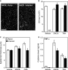
Dendritic cell migration, ear pathology, and TNF-α production associated with skin allergen exposure are regulated by a Heligmosomoides polygyrus bakeri infection. Naive and H. polygyrus bakeri-infected BALB/c mice were exposed to either the Th2-polarizing allergen Trimellitic anhydride (TMA), the Th1-polarizing allergen 2,4-dinitrochlorobenzene (DNCB), or vehicle. Whole ears were sectioned and stained to visualize tissue structure and allergen-associated epidermal thickening was measured (A). As an indicator of allergen-induced dendritic cells (DC) migration, the ear epidermal density of MHC class-II+ cells was ascertained, quantified (B), and visualized (C). ND = not done, scale bar = 50 μm. ALN cells were isolated and cultured, and supernatant levels of TNF-α were measured by Cytokine bead array (Becton Dickinson) (D). Values represent mean ± SE for four animals per group. * Significant difference between experimental groups (P < 0.05).

Similar articles

Cited by

References

    1. Maizels RM, et al. Immunological modulation and evasion by helminth parasites in human populations. Nature. 1993;365:797–805. - PubMed
    1. Anderson RM, May RM. Helminth infections of humans: mathematical models, population dynamics, and control. Adv Parasitol. 1985;24:1–101. - PubMed
    1. McSorley HJ, Loukas A. The immunology of human hookworm infections. Parasite Immunol. 2010;32:549–559. - PubMed
    1. Jackson JA, et al. T helper cell type 2 responsiveness predicts future susceptibility to gastrointestinal nematodes in humans. J Infect Dis. 2004;190:1804–1811. - PubMed
    1. Bell RG. IgE, allergies and helminth parasites: a new perspective on an old conundrum. Immunol Cell Biol. 1996;74:337–345. - PubMed

Publication types

MeSH terms

LinkOut - more resources