Randomized Controlled Trial
. 2014 Jul;2(7):616-31.
doi: 10.1158/2326-6066.CIR-14-0027. Epub 2014 Jun 18.

Immunotherapy converts nonimmunogenic pancreatic tumors into immunogenic foci of immune regulation

Affiliations
Randomized Controlled Trial

Immunotherapy converts nonimmunogenic pancreatic tumors into immunogenic foci of immune regulation

Eric R Lutz et al. Cancer Immunol Res. 2014 Jul.

Abstract

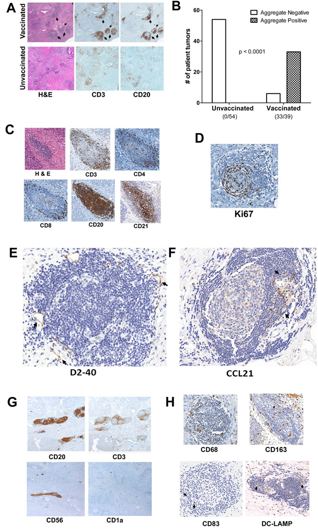
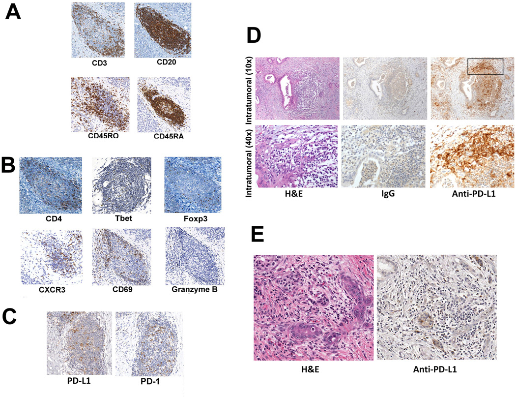
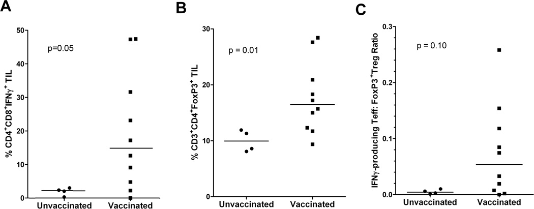
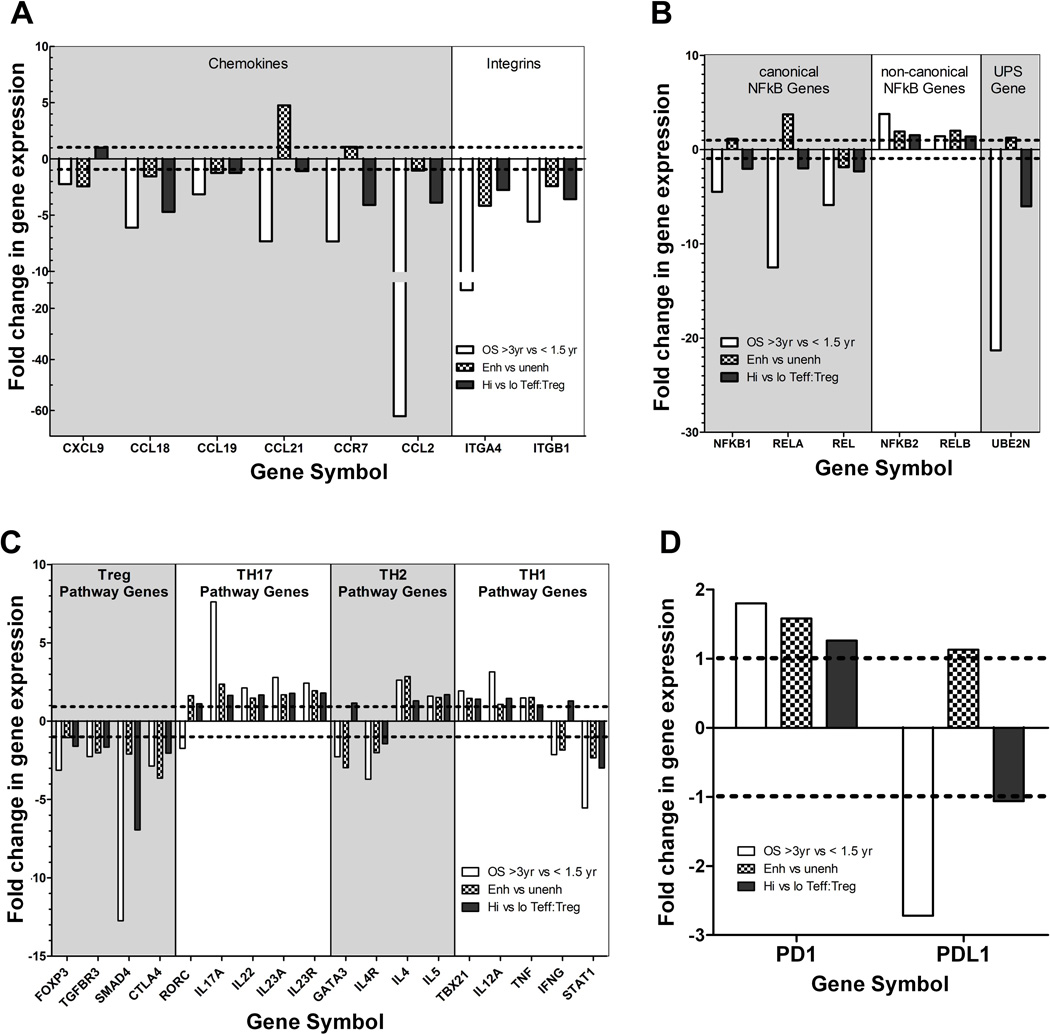
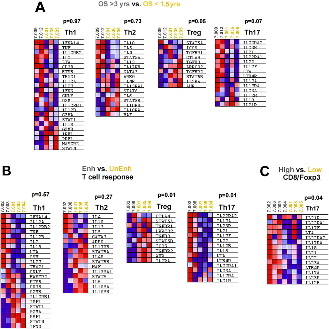
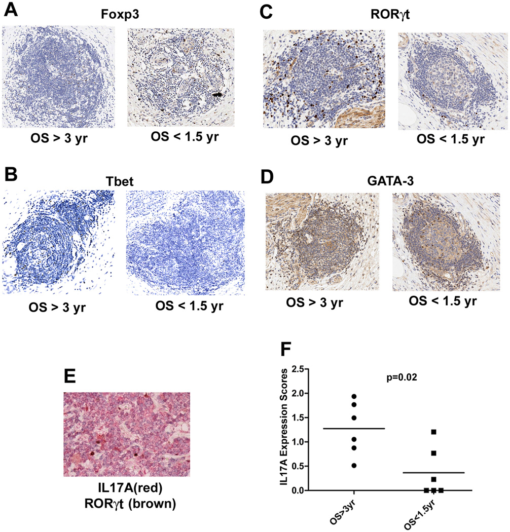
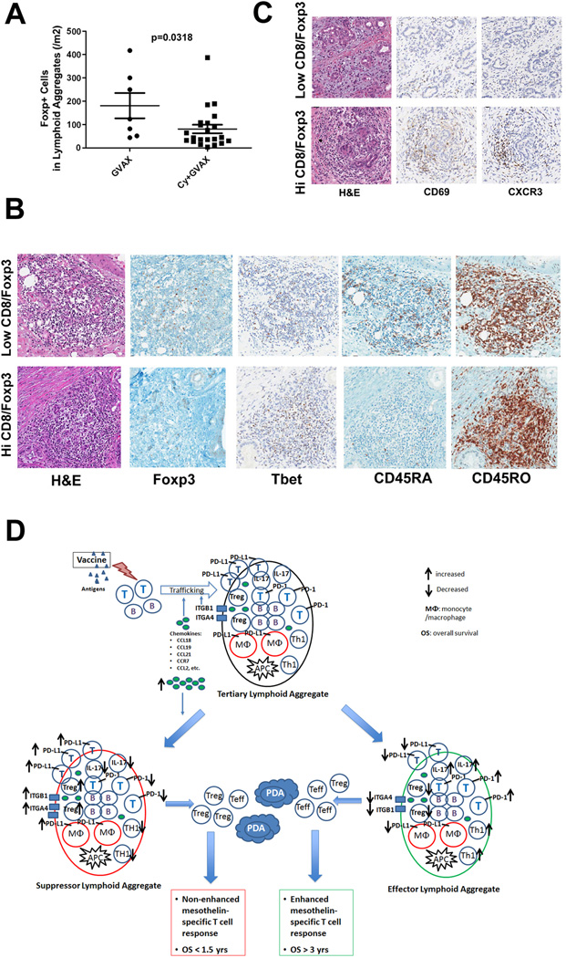
Pancreatic ductal adenocarcinoma (PDAC) is considered a "nonimmunogenic" neoplasm. Single-agent immunotherapies have failed to demonstrate significant clinical activity in PDAC and other "nonimmunogenic" tumors, in part due to a complex tumor microenvironment (TME) that provides a formidable barrier to immune infiltration and function. We designed a neoadjuvant and adjuvant clinical trial comparing an irradiated, granulocyte-macrophage colony-stimulating factor (GM-CSF)-secreting, allogeneic PDAC vaccine (GVAX) given as a single agent or in combination with low-dose cyclophosphamide to deplete regulatory T cells (Treg) as a means to study how the TME is altered by immunotherapy. Examination of resected PDACs revealed the formation of vaccine-induced intratumoral tertiary lymphoid aggregates in 33 of 39 patients 2 weeks after vaccine treatment. Immunohistochemical analysis showed these aggregates to be regulatory structures of adaptive immunity. Microarray analysis of microdissected aggregates identified gene-expression signatures in five signaling pathways involved in regulating immune-cell activation and trafficking that were associated with improved postvaccination responses. A suppressed Treg pathway and an enhanced Th17 pathway within these aggregates were associated with improved survival, enhanced postvaccination mesothelin-specific T-cell responses, and increased intratumoral Teff:Treg ratios. This study provides the first example of immune-based therapy converting a "nonimmunogenic" neoplasm into an "immunogenic" neoplasm by inducing infiltration of T cells and development of tertiary lymphoid structures in the TME. Post-GVAX T-cell infiltration and aggregate formation resulted in the upregulation of immunosuppressive regulatory mechanisms, including the PD-1-PD-L1 pathway, suggesting that patients with vaccine-primed PDAC may be better candidates than vaccine-naïve patients for immune checkpoint and other immunomodulatory therapies.


Conflict of interest statement

Conflict of interest:

The other authors have no conflict to disclose.

Figures

Figure 1
Figure 1. Intratumoral tertiary lymphoid aggregates observed in PDACs from Panc GVAX-treated patients
A. Lymphoid aggregates (indicated by arrows) observed in PDACs from Panc GVAX-treated (vaccinated) patients are not seen in Panc GVAX naïve (unvaccinated) patients. Lymphoid aggregates are composed of T cells (anti-CD3 staining) and B cells (anti-CD20 staining). B. Numbers of PDACs with (TLA+) or without (TLA) at least one lymphoid aggregate present in the intratumoral area in 54 unvaccinated patients and 39 vaccinated patients. Intratumoral lymphoid aggregates are observed specifically in PDACs from vaccinated patients (Fisher’s exact test p<0.001). C. IHC analysis with T-cell markers (CD3, CD4 and CD8), B-cell marker (CD20) and follicular dendritic cell marker (CD21). D. IHC analysis of the cell-proliferation marker Ki67. E. IHC analysis of the lymphatic-vessel (arrows) marker D2-40. F. IHC analysis of the chemokine ligand CCL21 (arrows). G. IHC analysis of CD20+ B cells, CD3+ T cells, CD56+ NK cells, and CD1a+ immature dendritic cells. Note that anti-CD56 staining also marks neural tissues (arrow). H. IHC analysis of CD68+ and CD163+ macrophages and CD83+ and DC-LAMP+ mature dendritic cells (arrows). All positive IHC signals are in brown.
Figure 2
Figure 2. Intratumoral lymphoid aggregates are sites of post-vaccination T-cell activation and regulation
A. IHC analysis of CD3-, CD20-, CD45RO-, and CD45RA-expressing cells. B. IHC analysis of CD4-, T-bet-, Foxp3-, CXCR3-, CD69-, and Granzyme B-expressing cells (Granzyme B+ cells are indicated by arrows). C. IHC analysis of B7-H1/PD-L1- and PD-1-expressing cells. D. PD-L1 expression in a representative intratumoral lymphoid aggregate from a vaccinated patient. The square indicates the edge of the intratumoral lymphoid aggregate that is shown in the bottom panels at a higher magnification. Brown staining indicates expression of the indicated markers. E. Absence of PD-L1 expression in a representative PDAC specimen from an unvaccinated patient.
Figure 3
Figure 3. Post-vaccination effector T-cell activation in intratumoral lymphoid aggregates is associated with Treg infiltration into the TME
Percentages of (A) IFNγ-producing CD4+CD8+ effector T cells and (B) CD3+CD4+FoxP3+ Tregs quantified by FACS in unstimulated TIL isolated from 10 vaccinated patients compared to those isolated from 3 unvaccinated patients. C. IFNγ-producing Teffector:Treg ratios measured in unstimulated TIL from the same sets of vaccinated and unvaccinated patients. Comparisons between vaccinated and unvaccinated TIL were made using Wilcoxon signed-rank tests and the calculated p values are shown.
Figure 4
Figure 4. Gene expression signatures in lymphoid aggregates associated with immune responses and overall survival
Gene expression measured by microarray was compared between microdissected lymphoid aggregates from 14 tumors grouped according to overall patient survival (OS > 3 years vs. < 1.5 years), post-vaccination induction of enhanced mesothelin-specific T-cell responses in PBL (Enh vs. Unenh), and the intratumoral CD8+ T effector/FoxP3+ Treg ratio (High vs. Low CD8/Foxp3 ratio). Gene ontology analysis revealed the enrichment of differentially expressed genes encoding numerous chemokine and chemokine receptor, integrin and adhesion molecule, NFkB pathway, and UPS components (Figure S6). Differences in Treg and Th17 pathway but not the Th1 or Th2 pathway genes were also associated with improved post-vaccination responses and survival (Figure S7). The linear fold-change in expression for each comparison is shown for representative genes in: (A) Chemokine, chemokine receptor, and integrin genes; (B) NFKB pathway genes; (C) T-helper pathway genes; and (D) PD-1 and PD-L1.
Figure 5
Figure 5. GSEA analyses of Th1, Th2, Th17 and Treg pathways
GSEA analyses were performed on the microarray data. A, Comparison between specimens from patients with OS > 3 years (subject numbers in grey) and patients with OS < 1.5 years (subject numbers in yellow); B, Comparison between specimens from patients who developed enhanced T-cell responses (in grey) and patients who did not (in yellow); C, Comparison between specimens from patients whose tumors were infiltrated with a high CD8/FoxP3 ratio (in grey) and whose tumors were infiltrated with a low CD8/FoxP3 ratio (in yellow). Heat maps of the microarray gene expression data are shown. Blue shading represents downregulation and red represents upregulation of gene expression. P values are shown for each GSEA comparison.
Figure 6
Figure 6. In situ expression of signature genes in the Th1, Th2, Th17 and Treg pathways
A. Anti-Foxp3 antibody staining for Tregs. B. Anti-T-bet staining for Th1 cells. C. Anti-RORγt staining for Th17 cells. D. Anti-GATA-3 staining for Th2 cells. For each target, one representative lymphoid aggregate from a patient with OS > 3 years and one from a patient with OS < 1.5 years is shown. All positive signals are in brown. E. Co-staining of IL17A (in red) and RORγt (in brown) in a representative lymphoid aggregate. F. Cumulative IL17 signals for all intratumoral lymphoid aggregates for each patient’s tumor section analyzed were quantified using Image Analysis Software (Aperio) and the expression levels were compared between patients with OS > 3 years (n=6) and OS < 1.5 years (n=6). Medians and the p value calculated using the Wilcoxon signed rank test are shown.
Figure 7
Figure 7. Low-dose cyclophosphamide reduces intratumoral Treg numbers and promotes enhanced T-cell trafficking and activation within the tertiary lymphoid aggregates
A. Comparison of the densities of Foxp3+ T cells in lymphoid aggregates between patients receiving GVAX alone (GVAX) and patients receiving GVAX plus Treg-modulating doses of Cy (CAX+Cy). B. IHC staining of Foxp3, T-bet, CD45RA, and CD45RO in representative lymphoid aggregates in PDACs from patients with high Teffector:Treg ratios in the TME compared to those with low Teffector:Treg ratios in the TME. C. IHC staining of CD69 and CXCR3 in representative PDAC tumor areas from patients with high versus low Teffector:Treg ratios in the TME. All positive IHC signals are in brown. D. Proposed model of vaccine-induced T-cell infiltration into the PDAC TME. At baseline, the “non-immunogenic” PDAC TME is primarily immunosuppressed and infiltrated with Tregs and other immunosuppressive populations of innate immune cells, and low numbers of effector T cells. Vaccination induces antigen-specific T cells systemically that traffic to the TME and initiate an inflammatory reaction. Chemokines including CCL2, CCL18, CCL19 and CCL21, and other signals produced during this inflammatory reaction recruit additional immune effector and regulatory cells, and induce the neogenesis of tertiary lymphoid aggregates within the TME, converting the PDAC TME into an “immunogenic” environment. These lymphoid aggregates are composed of effector T cells, B cells, Tregs and antigen-presenting cells (APCs), and serve as nodal sites for immune activation and regulation within the TME. Paradoxically, higher expression of the chemokines associated with the development of these aggregates favors the recruitment of immunosuppressive immune cells into the lymphoid aggregates in the TME. Lymphoid aggregates dominated by effector T cells and immune activation signals (“Effector Lymphoid Aggregates”) including: increased CD8+ effector T cells and effector T cells with Th17 signatures, increased PD-1 expression on effector T cells, decreased Tregs, and lower expression of PD-L1, are associated with higher Teffector:Treg ratios in TIL and enhanced post-vaccination T-cell responses in PBL, and generate productive antitumor responses that can prolong survival. In contrast, lymphoid aggregates dominated by immunosuppressive immune cells and signals (“Suppressor Lymphoid Aggregates”) including: decreased CD8+ effector T cells and effector T cells with Th17 signatures, decreased PD-1 expression on effector T cells, increased Tregs, and higher expression of PD-L1, are associated with decreased T effector:Treg ratios in TIL and unenhanced T-cell responses in PBL, do not generate productive antitumor responses, and cannot prolong survival. Use of targeted immune modulators may tip the balance between these signals in favor of the development of “effector lymphoid aggregates” and enhance the intratumoral immune responses induced by vaccines. However, without a vaccine, an “immunogenic” TME containing effector T cells and lymphoid aggregates is not available for immune modulators to act on.

References

    1. Society AC. Cancer Facts & Figures. Atlanta: 2012.
    1. Brahmer JR, Drake CG, Wollner I, Powderly JD, Picus J, Sharfman WH, et al. Phase I study of single-agent anti-programmed death-1 (MDX-1106) in refractory solid tumors: safety, clinical activity, pharmacodynamics, and immunologic correlates. J Clin Oncol. 2010;28:3167–3175. - PMC - PubMed
    1. Hodi FS, Butler M, Oble DA, Seiden MV, Haluska FG, Kruse A, et al. Immunologic and clinical effects of antibody blockade of cytotoxic T lymphocyte-associated antigen 4 in previously vaccinated cancer patients. Proc Natl Acad Sci U S A. 2008;105:3005–3010. - PMC - PubMed
    1. Sharma P, Wagner K, Wolchok JD, Allison JP. Novel cancer immunotherapy agents with survival benefit: recent successes and next steps. Nat Rev Cancer. 2011;11:805–812. - PMC - PubMed
    1. Clark CE, Beatty GL, Vonderheide RH. Immunosurveillance of pancreatic adenocarcinoma: insights from genetically engineered mouse models of cancer. Cancer Letters. 2009;279:1–7. - PubMed

Publication types

MeSH terms