. 2014 Oct;66(10):2881-91.
doi: 10.1002/art.38747.

Contribution of mast cell-derived interleukin-1β to uric acid crystal-induced acute arthritis in mice

Affiliations

Contribution of mast cell-derived interleukin-1β to uric acid crystal-induced acute arthritis in mice

Laurent L Reber et al. Arthritis Rheumatol. 2014 Oct.

Abstract

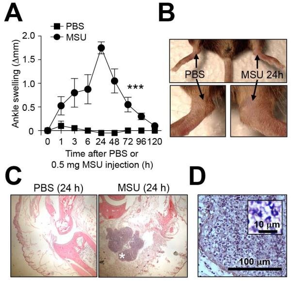
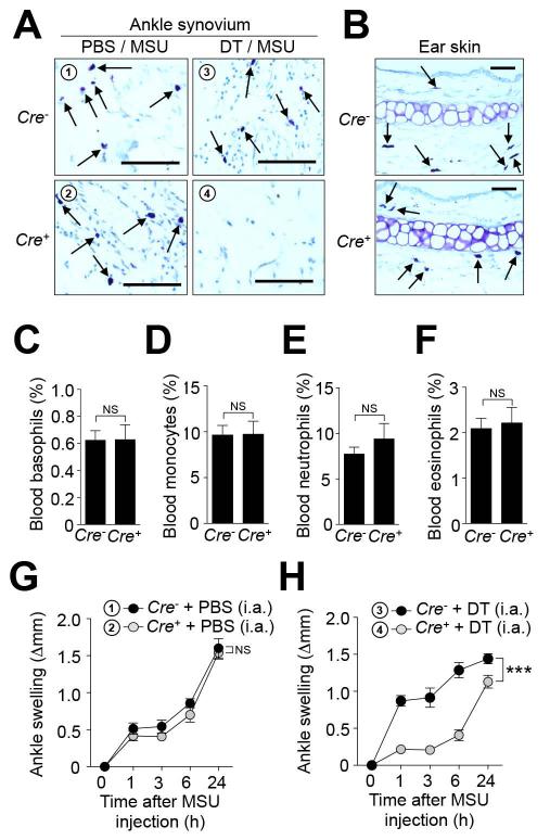
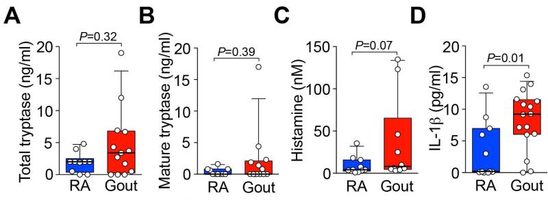
Objective: Gouty arthritis is caused by the precipitation of monosodium urate monohydrate (MSU) crystals in the joints. While it has been reported that mast cells (MCs) infiltrate gouty tophi, little is known about the actual roles of MCs during acute attacks of gout. This study was undertaken to assess the role of MCs in a mouse model of MSU crystal-induced acute arthritis.

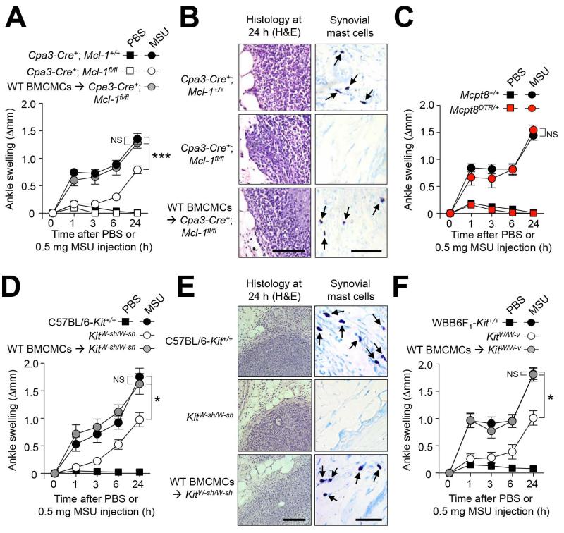
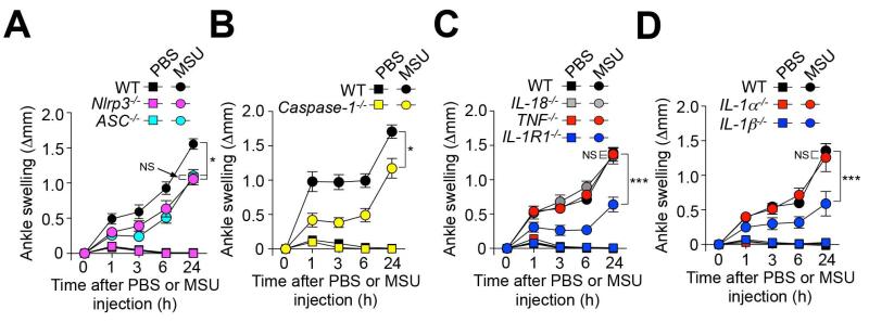
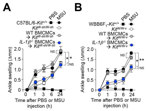
Methods: We assessed the effects of intraarticular (IA) injection of MSU crystals in various strains of mice with constitutive or inducible MC deficiency or in mice lacking interleukin-1β (IL-1β) or other elements of innate immunity. We also assessed the response to IA injection of MSU crystals in genetically MC-deficient mice after IA engraftment of wild-type or IL-1β(-/-) bone marrow-derived cultured MCs.

Results: MCs were found to augment acute tissue swelling following IA injection of MSU crystals in mice. IL-1β production by MCs contributed importantly to MSU crystal-induced tissue swelling, particularly during its early stages. Selective depletion of synovial MCs was able to diminish MSU crystal-induced acute inflammation in the joints.

Conclusion: Our findings identify a previously unrecognized role of MCs and MC-derived IL-1β in the early stages of MSU crystal-induced acute arthritis in mice.


Figures

Figure 1
Figure 1. Mouse model of MSU-induced acute arthritis
C57BL/6J mice were injected i.a. with MSU crystals (0.5 mg in 10 μl) in one ankle and vehicle (10 μl PBS) in the contra-lateral ankle. (A and B) Time course and representative photographs (at 24 h) of MSU-induced ankle swelling. (C) H&E-stained sections of ankles at 24 h. (D) Enlargement of the area marked (*) in (C). Insert in (D) shows enlargement of leukocyte infiltrate. Data in (A) are means ± SEM from two independent experiments. *** = P < 0.001 vs. indicated groups by ANOVA. Scale bar in D: 100 μm (insert in D: 10 μm).
Figure 2
Figure 2. MCs can amplify MSU crystal-induced ankle swelling
(A, C, D, F) Changes in ankle thickness after i.a injection of 0.5 mg MSU or PBS in (A) MC- and basophil-deficient Cpa3-Cre+; Mcl-1fl/fl (n=17), Cpa3-Cre+; Mcl-1+/+ littermate (n=20) and C57BL/6J (WT) BMCMC i.a. engrafted Cpa3-Cre+; Mcl-1fl/fl mice (n=10), (C) DT-treated basophil-deficient Mcpt8DTR/+ (n=9) and Mcpt8+/+ littermate (n=9) mice, (D) C57BL/6-Kit+/+ (n=13), MC-deficient KitW-sh/W-sh (n=12), and C57BL/6J (WT) BMCMC i.a. engrafted KitW-sh/W-sh (n=12) mice, and (F) WBB6F1-Kit+/+ (WT) (n=10), MC-deficient WBB6F1-KitW/W-v (n=10) and WBB6F1-Kit+/+ (WT) BMCMC i.a. engrafted WBB6F1-KitW/W-v (n=10) mice. (B and E) H&E- (for leukocytes) and Toluidine blue- (for MCs) stained sections of ankles at 24 h. (A, C, D, F) Data are means ± SEM from three (C, D and F) or three to five (A) independent experiments. * or *** = P < 0.05 or 0.001 vs. indicated groups by ANOVA. Scale bars: 100 μm. NS, not significant (P > 0.05).
Figure 3
Figure 3. Contributions of the NLRP3 inflammasome, IL-1R1, and IL-1β to MSU crystal-induced ankle swelling
Changes in ankle thickness in (A) C57BL/6J (WT) (n=11), Nlrp3−/− (n=9) and ASC−/− (n=10) mice; (B) C57BL/6J (WT) (n=14) and Caspase-1−/− (n=11) mice; (C) C57BL/6J (WT) (n=16), TNF−/− (n=12), IL-18−/− (n=10) and IL-1R1−/− (n=13) mice; and (D) C57BL/6J (WT) (n=9), IL-1α−/− (n=7) and IL-1β−/− (n=10) after i.a. injection of 0.5 mg MSU or PBS. Data are shown as means ± SEM. * or *** = P < 0.05 or 0.001 vs. indicated groups by ANOVA. Differences in swelling between MSU crystal-injected vs. the corresponding PBS-injected ankles are significant at each time point (P < 0.05 by unpaired Student’s t-test) for all groups of mice. NS, not significant (P > 0.05).
Figure 4
Figure 4. Contributions of MC-derived IL-1β to MSU crystal-induced ankle swelling
Changes in ankle thickness after i.a injection of 0.5 mg MSU or PBS in (A) C57BL/6-Kit+/+ (n=8) or MC-deficient KitW-sh/W-sh mice (n=6), and KitW-sh/W-sh mice engrafted i.a. with C57BL/6J (WT) (n=11) or C57BL6-IL-1β−/− (n=10) BMCMCs; and (B) WBB6F1-Kit+/+ (n=17) or MC-deficient WBB6F1-KitW/W-v mice (n=8), and WBB6F1-KitW/W-v mice engrafted i.a. with C57BL/6J (WT) (n=15) or C57BL/6-IL-1β−/− (n=11) BMCMCs. Data are shown as means ± SEM from three independent experiments (except for MC-deficient KitW-sh/W-sh mice, which were included in two of the three independent experiments). * or ** = P < 0.05 or 0.01 vs. indicated groups by ANOVA. NS, not significant (P > 0.05).
Figure 5
Figure 5. Local and selective ablation of MCs reduces MSU crystal-induced ankle swelling
Cpa3-Cre+; DTRfl/+ (Cre+; n=13) and Cpa3-Cre; DTRfl/+ (Cre; n=7) mice were injected i.a. with diphtheria toxin (DT) (two successive weekly injections of 50 ng) in one ankle and vehicle (PBS) in the contralateral ankle. 0.5 mg MSU was injected into both ankles 1 week after the last DT injection. (A and B) Toluidine-blue staining of ankle joint tissue (A) showing ablation of synovial MCs after treatment with DT (but not PBS) in Cre+ mice and the presence of MCs in ear skin (B) in either Cre or Cre+ mice. Scale bars: 50 μm. (C-F) Blood leukocytes isolated 1 h before MSU injection were analyzed by flow cytometry. (C-F) % (mean + SEM) of (C) basophils (CD49b+; IgE+), (D) monocytes (Gr-1low; CD11b+; Siglec-F), (E) neutrophils (Gr-1high; CD11b+; Siglec-F) & (F) eosinophils (SSChigh; Siglec-F+). NS, not significant (P > 0.05 by unpaired Student’s t test). (G and H) Changes in ankle thickness (means ± SEM) after i.a injection of MSU. Data are shown as means ± SEM from two (for Cre mice) or three (for Cre+ mice) independent experiments. *** = P < 0.001 vs. indicated group by ANOVA.
Figure 6
Figure 6. Levels of tryptase, histamine and IL-1β in synovial fluids from patients with gout or rheumatoid arthritis (RA)
Levels of total (A) and mature (B) tryptase, histamine (C) and IL-1β (D) measured by ELISA in synovial fluids from patients with RA (n=10-11) or gout (n=10-16). Data are represented as box and whisker plots with bottom and top of the boxes showing the 25th & 75th percentiles and bars showing the 10th & 90th percentiles; individual data are plotted as circles. P values were calculated using a Mann-Whitney test.

References

    1. Lawrence RC, Felson DT, Helmick CG, Arnold LM, Choi H, Deyo RA, et al. Estimates of the prevalence of arthritis and other rheumatic conditions in the United States. Part II. Arthritis Rheum. 2008;58(1):26–35. - PMC - PubMed
    1. Martinon F, Petrilli V, Mayor A, Tardivel A, Tschopp J. Gout-associated uric acid crystals activate the NALP3 inflammasome. Nature. 2006;440(7081):237–41. - PubMed
    1. Chen CJ, Shi Y, Hearn A, Fitzgerald K, Golenbock D, Reed G, et al. MyD88-dependent IL-1 receptor signaling is essential for gouty inflammation stimulated by monosodium urate crystals. J Clin Invest. 2006;116(8):2262–71. - PMC - PubMed
    1. Abraham SN, St John AL. Mast cell-orchestrated immunity to pathogens. Nat Rev Immunol. 2010;10(6):440–52. - PMC - PubMed
    1. Nigrovic PA, Binstadt BA, Monach PA, Johnsen A, Gurish M, Iwakura Y, et al. Mast cells contribute to initiation of autoantibody-mediated arthritis via IL-1. Proc Natl Acad Sci U S A. 2007;104(7):2325–30. - PMC - PubMed

Publication types

LinkOut - more resources