. 2014 Jun 18;5(3):79.
doi: 10.1186/scrt468.

Diabetes impairs the angiogenic potential of adipose-derived stem cells by selectively depleting cellular subpopulations

Diabetes impairs the angiogenic potential of adipose-derived stem cells by selectively depleting cellular subpopulations

Robert C Rennert et al. Stem Cell Res Ther. .

Abstract

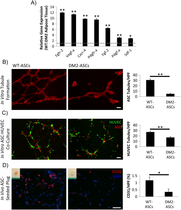
Introduction: Pathophysiologic changes associated with diabetes impair new blood vessel formation and wound healing. Mesenchymal stem cells derived from adipose tissue (ASCs) have been used clinically to promote healing, although it remains unclear whether diabetes impairs their functional and therapeutic capacity.

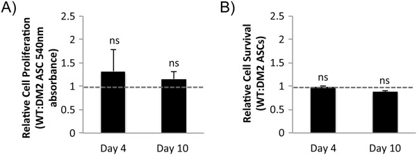
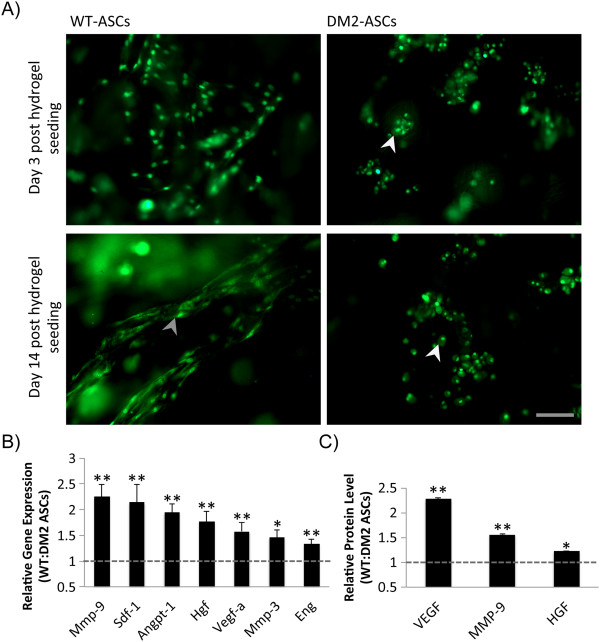
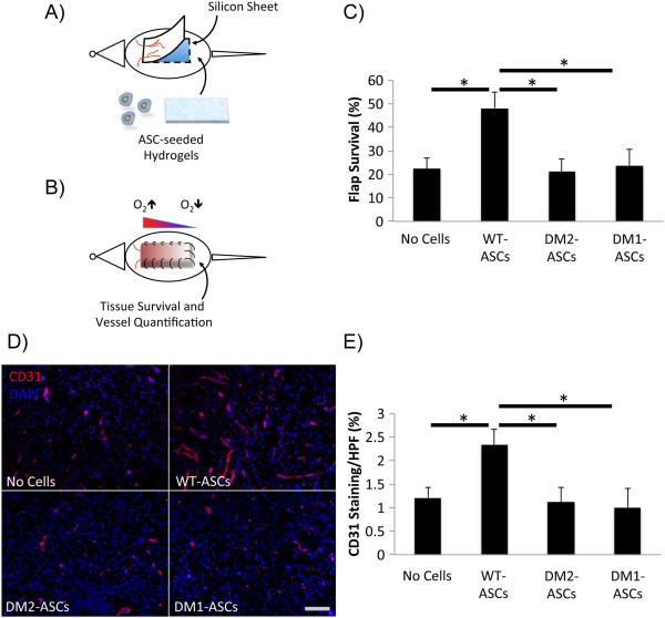
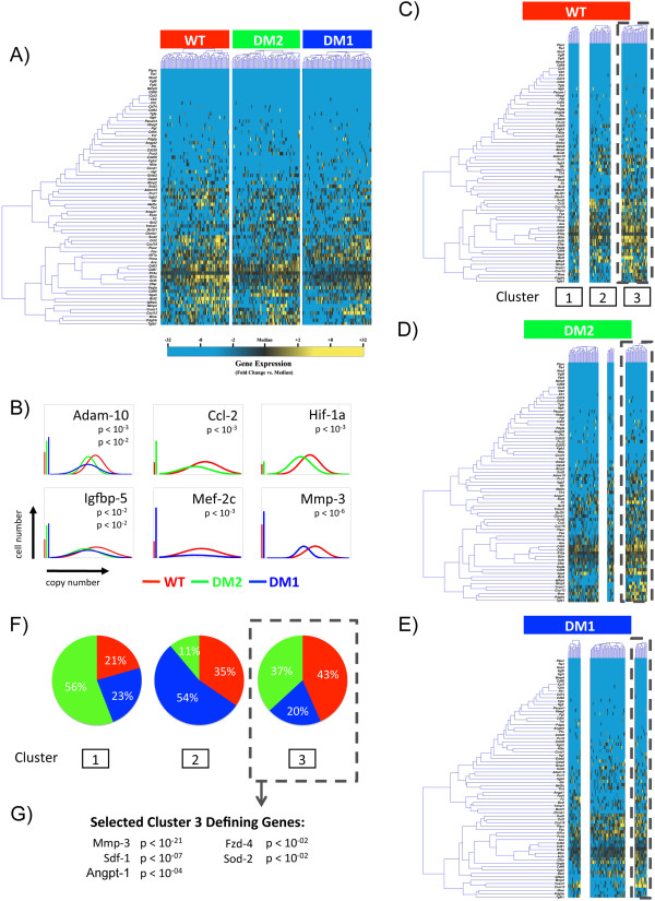
Methods: In this study, we examined the impact of diabetes on the murine ASC niche as well as on the potential of isolated cells to promote neovascularization in vitro and in vivo. A novel single-cell analytical approach was used to interrogate ASC heterogeneity and subpopulation dynamics in this pathologic setting.

Results: Our results demonstrate that diabetes alters the ASC niche in situ and that diabetic ASCs are compromised in their ability to establish a vascular network both in vitro and in vivo. Moreover, these diabetic cells were ineffective in promoting soft tissue neovascularization and wound healing. Single-cell transcriptional analysis identified a subpopulation of cells which was diminished in both type 1 and type 2 models of diabetes. These cells were characterized by the high expression of genes known to be important for new blood vessel growth.

Conclusions: Perturbations in specific cellular subpopulations, visible only on a single-cell level, represent a previously unreported mechanism for the dysfunction of diabetic ASCs. These data suggest that the utility of autologous ASCs for cell-based therapies in patients with diabetes may be limited and that interventions to improve cell function before application are warranted.


Figures

Figure 1
Figure 1
Adipose-derived mesenchymal stem cell (ASC) niche characterization and in vitro and in vivo analyses of ASC neovascular potential. (A) Transcriptional profile of type 2 diabetes mellitus (DM2) and wild-type (WT) murine fat pads. A dysfunctional in situ signaling environment was observed in the setting of diabetes. (B) Matrigel culture of WT and DM2 ASCs under hypoxic conditions. Tubules per high-power field (HPF) were quantified as a surrogate for direct ASC vasculogenic potential. (C) Matrigel co-culture of ASCs and human umbilical vein endothelial cells (HUVECs) under hypoxic conditions. HUVEC tubules per HPF were quantified as a measure of ASC-mediated endothelial network formation. (D) CD31 staining to quantify in vivo Matrigel plug vascularity following seeding with either WT or DM2 ASCs. Insets provide gross images of explanted plugs. Scale bar = 50 μm. *P ≤0.05, **P <0.01.
Figure 2
Figure 2
Adipose-derived mesenchymal stem cell (ASC) adipogenic and osteogenic differentiation. (A) Representative images and (B) quantification of Oil red O and Alizarin red staining following adipogenic and osteogenic differentiation of WT and DM2 ASCs. Scale bar = 50 μm. **P <0.01.
Figure 3
Figure 3
ASC behavior following in vitro hydrogel seeding. (A,B) Wild-type (WT) and diabetic (DM2) ASCs display similar rates of (A) proliferation and (B) survival in vitro following seeding within a three-dimensional bioscaffold. ns, not significant (P >0.05).
Figure 4
Figure 4
Adipose-derived mesenchymal stem cell (ASC) incorporation and secretory function following hydrogel bioscaffold seeding. (A) Wild-type (WT) and type 2 diabetes mellitus (DM2) ASCs at day 3 and 14 after hydrogel seeding. WT cells showed a uniform incorporation within the hydrogel and developed cell elongations and cytoplasmic extensions indicative of interactions with the bioscaffold (gray arrowhead). DM2 ASCs displayed a clumped distribution with rounded cell morphology (white arrowheads). (B) Reverse transcription-polymerase chain reaction and protein array (C) quantifying the relative expression of selected genes associated with vasculogenenesis and tissue remodeling in hydrogel-seeded WT versus DM2 ASCs. Scale bar = 50 μm. *P ≤ 0.05, **P <0.01. Angpt-1, angiopoietin 1; Eng, endoglin; Hgf, hepatocyte growth factor; Mmp-3, matrix metalloproteinase 3; Mmp-9, matrix metalloproteinase 9; Sdf-1, Stromal Cell-Derived Factor 1; Vegf, vascular endothelial cell growth factor.
Figure 5
Figure 5
Hydrogel delivery of ASCs to promote ischemic tissue flap survival. (A) WT and DM (1 and 2) ASC-seeded hydrogels and unseeded controls were inset following creation of a full-thickness peninsular skin flap, (B) resulting in the creation of a reproducible ischemic gradient. (C) Tissue survival and vascularization as determined by (D) CD31 immunohistochemistry staining and (E) quantification were assessed at day 10. Scale bar = 50 μm. *P ≤0.05.
Figure 6
Figure 6
Diabetic and wild-type (WT) stromal vascular fraction (SVF) cell surface marker analysis. (A) Flow cytometric analysis determining the percentage of putative adipose-derived mesenchymal stem cells (ASCs) (CD45-/31-/34+ cells) within SVF obtained from diabetic (DM2 and DM1) and WT mice (CD45 and live/dead gating not shown). (B) Quantification of CD45-/31-/34+ ASCs in WT, DM2, and DM1 SVF reveals a significant depletion of ASCs in diabetic samples. **P ≤0.01.
Figure 7
Figure 7
Single-cell transcriptional analysis of CD45-/CD31-/CD34+ ASCs. (A) Hierarchical clustering of cells from wild-type (WT; left), db/db diabetic (DM2; middle), and STZ-induced diabetic (DM1; right) mice. Gene expression is presented as fold change from median on a color scale from yellow (high expression, 32-fold above median) to blue (low expression, 32-fold below median). See Additional file 2: Figure S1 for complete dataset. (B) Differentially expressed genes between wild-type (WT) and diabetic (db/db [DM2] or STZ [DM1]) cells identified by using non-parametric two-sample Kolmogorov-Smirnov testing. Twenty-one genes exhibited significantly different (P <0.01 following Bonferroni correction for multiple comparisons) distributions of single-cell expression between populations; six are illustrated here by using median-centered Gaussian curve fits. The left bar for each panel represents the fraction of qPCR that failed to amplify in each group. Curves and p values for each gene are shown only for those diabetic groups significantly different from wild-type cells. (C) wild-type, (D) DM2, and (E) DM1 cells based on the expression patterns of all 71 genes (k = 3). (F) Pie charts representing the fraction of ASCs comprising each cluster (WT [red], DM2 [green], and DM1 [blue]). (G) List of selected cluster 3-defining genes.

Comment in

References

    1. Brem H, Tomic-Canic M. Cellular and molecular basis of wound healing in diabetes. J Clin Invest. 2007;117:1219–1222. - PMC - PubMed
    1. Tepper OM, Galiano RD, Capla JM, Kalka C, Gagne PJ, Jacobowitz GR, Levine JP, Gurtner GC. Human endothelial progenitor cells from type II diabetics exhibit impaired proliferation, adhesion, and incorporation into vascular structures. Circulation. 2002;106:2781–2786. - PubMed
    1. Lerman OZ, Galiano RD, Armour M, Levine JP, Gurtner GC. Cellular dysfunction in the diabetic fibroblast: impairment in migration, vascular endothelial growth factor production, and response to hypoxia. Am J Pathol. 2003;162:303–312. - PMC - PubMed
    1. Thangarajah H, Yao D, Chang EI, Shi Y, Jazayeri L, Vial IN, Galiano RD, Du XL, Grogan R, Galvez MG, Januszyk M, Brownlee M, Gurtner GC. The molecular basis for impaired hypoxia-induced VEGF expression in diabetic tissues. Proc Natl Acad Sci U S A. 2009;106:13505–13510. - PMC - PubMed
    1. Thangarajah H, Vial IN, Grogan RH, Yao D, Shi Y, Januszyk M, Galiano RD, Chang EI, Galvez MG, Glotzbach JP, Wong VW, Brownlee M, Gurtner GC. HIF-1alpha dysfunction in diabetes. Cell Cycle. 2010;9:75–79. - PubMed

Publication types