. 2014 Sep 15;307(6):R608-20.
doi: 10.1152/ajpregu.00085.2014. Epub 2014 Jun 18.

Differential coupling of KLF10 to Sin3-HDAC and PCAF regulates the inducibility of the FOXP3 gene

Affiliations

Differential coupling of KLF10 to Sin3-HDAC and PCAF regulates the inducibility of the FOXP3 gene

Yuning Xiong et al. Am J Physiol Regul Integr Comp Physiol. .

Abstract

Inducible gene expression, which requires chromatin remodeling on gene promoters, underlies the epigenetically inherited differentiation program of most immune cells. However, chromatin-mediated mechanisms that underlie these events in T regulatory cells remain to be fully characterized. Here, we report that inducibility of FOXP3, a key transcription factor for the development of T regulatory cells, depends upon Kruppel-like factor 10 (KLF10) interacting with two antagonistic histone-modifying systems. We utilized chromatin immunoprecipitation, genome-integrated reporter assays, and functional domain KLF10 mutant proteins, to characterize reciprocal interactions between this transcription factor and either the Sin3-histone deacetylase complex or the histone acetyltransferase, p300/CBP-associated factor (PCAF). We characterize a Sin3-interacting repressor domain on the NH2 terminus of KLF10, which works to limit the activating function of this transcription factor. Indeed, inactivation of this Sin3-interacting domain renders KLF10 able to physically associate with PCAF as to induce FOXP3 gene transcription. We show that this biochemical data derived from studying our genome-integrated reporter cell system are recapitulated in primary murine lymphocytes. Collectively, these results advance our understanding of how a single transcription factor, namely KLF10, functions as a toggle to integrate antagonistic signals regulating FOXP3 and, thus, immune activation.

Keywords: FOXP3; KLF10; PCAF; Sin3; T regulatory cell.


Figures

Fig. 1.
Fig. 1.
Kruppel-like factor 10 (KLF10) represses the core FOXP3 promoter in vitro. Luciferase reporter assays in the genome-integrated Jurkat cell lines demonstrating repression of core FOXP3 promoter function by KLF10. Relative luciferase units (RLU) upon overexpression of KLF family members into FLP-core cells with (A) or without (B) activating conditions (see materials and methods). KLF10 uniquely represses FOXP3 promoter function in both activating and resting conditions (0.63 ± 0.04 and 0.53 ± 0.10 RLU, second column Fig. 1, A and B, respectively). RLUs are normalized to EV control (1.00, white column). The data are expressed as means ± SE of three independent experiments with *P = 0.035 (A) and P = 0.004 (B). C: chromatin immunoprecipitation assay demonstrating binding of KLF10 to the FOXP3 core promoter locus. Inset: representative DNA gel for PCR reaction analysis of the expression of FOXP3 in cell fractions postimmunoprecipitation for His. Quantitative real-time PCR analysis of the expression of FOXP3 in cell fractions postimmunoprecipitation for His-tagged KLF10 in FLP-core cells transfected with KLF10-His expression vector demonstrates significant binding of KLF10 to the core promoter (3.81 ± 0.70-fold change over EV control). Results are presented controlled to FOXP3 expression of preimmunoprecipitated sample (input). EV, empty vector. The data are representative of three independent experiments with *P = 0.04.
Fig. 2.
Fig. 2.
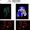
Putative structural model for the Sin3-PAH2 and KLF10-Sin3 interacting domain (SID) complex. Molecular modeling of KLF10 and Sin3 interaction utilizing HBP1 as a template was performed to predict disruptive domain mutations. A: sequence alignment of HBP1 SID, putative wild-type (wt) KLF10 SID, and E5P, A6P double KLF10 SID mutant. The established SID HBP1 was used as our modeling template. Consensus of the minimum SID is demonstrated in shaded gray. B: upper apolar surface of KLF10 SID alpha helix established hydrophobic interactions within the similarly charged pocket created by the four-helix bundle of the Sin3-PAH2. C: sequestration of hydrophobic sidechains (depicted as brown) away from hydrophilic surface (depicted as blue) to form the hydrophobic pocket, which accommodates the KLF10 SID. D: critical bonds predicted between KLF10SID and Sin3 upon examination of protein-protein interaction interphase. Pairs of interacting residues are labeled with the same color, while bonds are indicated by lines. E: predicted disruption of binding between KLF10 SID E5 with Sin3 H333 and KLF10 A6 with Sin3 V311 by proline mutagenesis. Pairs of interacting residues are labeled with the same color, while bonds are indicated by lines. Note the disappearance of lines indicating disruption of bonds upon mutations of E5 and A6 to P.
Fig. 3.
Fig. 3.
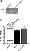
Proline mutagenesis of the SID disrupts KLF10-Sin3 protein/protein interaction but not KLF10 DNA binding. A: immunoblot using antibody specific for Sin3 (top) or His tag (bottom) of Jurkat cell lysates after overexpression of His-tagged KLF10 wt (left lane) or KLF10 SID mutant construct (right lane). Note that as predicted in the molecular modeling experiments, wt KLF10 physically interacts with Sin3, and the proline mutagenesis completely disrupts this interaction. The data are representative of three independent experiments. B: KLF10 or KLF10 SID binding mutant constructs were expressed in Jurkat cells to demonstrate by chromatin immunoprecipitation the preserved competence of DNA binding by the KLF10 SID mutant construct. Inset, top: representative DNA gel for PCR reaction analysis of the expression of FOXP3 in cell fractions postimmunoprecipitation for His. Bottom: quantitative real-time PCR analysis of the expression of FOXP3 in cell fractions postimmunoprecipitation for His-tagged KLF10 demonstrates significant binding of both KLF10 (black column, 3.63 ± 0.3-fold change) and KLF10-SIDmt (gray column, 4.29 ± 0.2-fold change) constructs to the core promoter compared with EV control. Note the ability of the KLF10-SID mutant to bind DNA, equivalent to wt KLF10. The data are representative of three independent experiments; *P = 0.024.
Fig. 4.
Fig. 4.
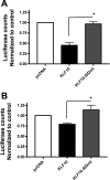
KLF10 represses FOXP3 through association with Sin3. Luciferase reporter assays in genome-integrated Jurkat cell lines demonstrating disruption of repressor function upon mutation of the KLF10 SID. Relative luciferase counts upon overexpression of KLF10 or KLF10-SIDmt into FLP core (A) or FLP-core E1 cells (B), normalized to empty vector control (1.00, white column). Note the established repression of FOXP3 gene transcription by wt KLF10 is abrogated by proline mutagenesis of the KLF10-SID [0.97 ± 0.05 vs. 0.45 ± 0.07 RLU (A); and 1.14 ± 0.12 vs. 0.79 ± 0.03 RLU (B)]. The data are expressed as means ± SE of six independent experiments, *P = 0.003 (A); *P = 0.009 (B).
Fig. 5.
Fig. 5.
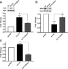
KLF10 recruits HDAC chromatin-modifying complexes to the FOXP3 promoter. Chromatin immunoprecipitation assay demonstrating that recruitment of the Sin3-HDAC repressor complex to the FOXP3 core promoter locus is dependent upon the KLF10 SID. Quantitative real-time PCR analysis of the expression of FOXP3 in cell fractions postimmunoprecipitation for Sin3 (A), H4 polyacetylation (B), or HDAC1 (C) in FLP-core-E1 cells transfected with KLF10 or KLF10-SIDmt expression vectors. Note the capacity of wt KLF10 but not the KLF10-SIDmt protein to recruit Sin3 (2.717 ± 0.35 vs. 1.63 ± 0.09-fold change; *P = 0.024) and histone deacetylase HDAC1 (1.25 ± 0.12-fold change vs. 0.43 ± 0.05-fold change; *P = 0.024) to the core promoter resulting in loss of baseline histone 4 acetylation state (0.44 ± 0.15 vs. 0.82 ± 0.14-fold change; *P = 0.034). The data represent means ± SE of three independent experiments. Inset: representative DNA gel for PCR reaction analysis of the expression of FOXP3 in cell fractions postimmunoprecipitation for Sin3 (A) and poly H4-Ac (B).
Fig. 6.
Fig. 6.
Derepression depends upon the histone acetyltransferase (HAT), p300/CBP-associated factor (PCAF). Luciferase reporter assay demonstrates that abrogation of repressor function evident upon mutation of the KLF10 SID is dependent upon the HAT, PCAF. Relative luciferase counts, normalized to empty vector control (1.00; white column), upon overexpression of KLF10 (black columns), or KLF10-SIDmt (columns 3–5) into FLP-core cells with concomitant knockdown of PCAF (PCAF siRNA) or scramble siRNA (scr) control. KLF10 represses FOXP3 promoter function [0.62 ± 0.03 relative light units (RLU), second column from the left] only when the SID domain is intact (1.65 ± 0.34 RLU KLF10-SIDmt, second column from the right). Note the derepression evident upon proline mutagenesis of the KLF10-SIDmt (second column from the right) entirely depends upon the expression of PCAF (1.65 ± 0.34 RLU, second column from the right vs. 0.58 ± 0.13 RLU, furthest right column; P = 0.005). The data express the means ± SE of 3 independent experiments.
Fig. 7.
Fig. 7.
KLF10 associates with PCAF. Assays of protein-protein interaction demonstrate physiologically relevant interaction between PCAF and the N-terminus domain of KLF10. A: immunoblot for PCAF in cell-free association assay of purified PCAF and GST constructs indicated. Note in this cell-free assay, PCAF associates with both the NH2 and COOH terminus of KLF10. B: immunoblot for PCAF using lysates of Jurkat lymphocytes incubated with GST fusion proteins (GST control, GST-KLF10 1–210, or GST-KLF10 210–350). Purified PCAF run on the left lane represents the positive control. Note that in a physiologically relevant system using Jurkat cell lysates, PCAF clearly interacts with the NH2 terminus, but not the COOH terminus of KLF10.
Fig. 8.
Fig. 8.
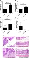
Rag2−/− mice reconstituted with KLF10−/− bone marrow exhibit more severe colitis than Rag2−/− mice reconstituted with wt bone marrow (BM). Data are presented as means ± SE (n = 6 mice per group). A: weight loss at the time of death. The values of body weight are expressed as a percentage of initial body weight on day 0. Rag2−/− reconstituted with KLF10−/− BM demonstrated significantly enhanced weight loss compared with mice reconstituted with wt BM (6.89 ± 0.53 vs. 3.64 ± 1.31%, P = 0.04). B: macroscopic disease score based on the presence of clinical signs of colitis (wasting and hunching of the recipient mouse and the physical characteristics of stool) and an ordinal scale of colonic involvement (thickness and erythema) shows Rag2−/− reconstituted with KLF10−/− BM had significantly enhanced disease activity compared with mice reconstituted with Wt lymphocytes (4.31 ± 0.38 vs. 2.64 ± 0.33; P = 0.01). Levels of cytokines IL-6 (C) and IL-10 (D) were determined in serum using mouse cytometric bead array cytokine assay (BD Biosciences, San Jose, CA, USA) and analyzed using FCAP array version 3 software (Soft Flow Hungary). Rag2−/− reconstituted with KLF10−/− lymphocytes demonstrated higher levels of proinflammatory cytokine IL-6 (166.3 ± 41.34 vs. 65.65 ± 27.60 pg/ml; P = 0.11) and lower levels of anti-inflammatory cytokine IL-10 than Rag2−/− mice reconstituted with Wt bone marrow (0.56 ± 0.56 vs. 6.60 ± 3.82 pg/ml; P = 0.08). E: representative histologic sections of mouse colon upon death from the two experimental conditions. Note a higher degree of crypt loss and diffuse eosinophilic infiltration in the KLF10−/− recipient animals compared with WT recipient controls.
Fig. 9.
Fig. 9.
Quantitative real-time PCR analysis of the expression of KLF10 in primary naïve CD4+ murine lymphocyte cells transduced with KLF10 or KLF10-SIDmt expression vectors. Note that overexpression of KLF10 adenoviral vectors results in 5–10-fold increased expression over the empty adenoviral vector. Results are presented controlled to KLF10 expression of empty vector-transduced cells.
Fig. 10.
Fig. 10.
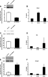
: KLF10 associates alternatively with Sin3 or PCAF to repress or activate FOXP3 gene transcription in primary naïve CD4+ lymphocytes. Using adenoviral constructs in primary murine lymphocytes, chromatin immunoprecipitation demonstrates recruitment of alternatively Sin 3 or PCAF to the FOXP3 core promoter locus to depend upon the KLF10 SID. A, C, E: Inset: DNA gel for PCR reaction analysis of the expression of FOXP3 in cell fractions postimmunoprecipitation for Sin3 (A), poly histone 4 (H4)-Ac (C), and PCAF (C) in primary lymphocytes posttransduction with KLF10 or KLF10-SIDmt expression vectors. Histogram represents densitometry units normalized by input for associated gels. Note that recapitulating the data from cell lines, wt KLF10, but not the KLF10-SIDmt protein, recruits Sin3 to the core promoter (2.43 ± 0.18 vs. 1.00 ± 0.1 densitometry units normalized to input), resulting in a decrease of the histone 4 acetylation state (52.45 ± 4.45 vs. 77.95 ± 7.14 densitometry units normalized to input; C). KLF10-SIDmt recruits PCAF to the core promoter (11.43 ± 1.58 vs. 2.51 ± 1.09 densitometry units normalized to input). The data are expressed as means ± SE of three independent experiments. B, D, F: quantitative real-time PCR analysis of the expression of FOXP3 in cell fractions postimmunoprecipitation for Sin3 (B), H4 polyacetylation (D), or PCAF (F) in primary T cells transfected with EV, KLF10, or KLF10-SIDmt expression vectors. Note the capacity of wt KLF10 but not the KLF10-SIDmt protein to recruit Sin3 (2.93 ± 0.25 vs. 1.23 ± 0.30-fold change; P = 0.04). In the absence of Sin3, the KLF10-SIDmt recruits PCAF (4.56 ± 1.22-fold change vs. 0.51 ± 0.03-fold change) to the core promoter resulting in enhancement of the histone 4 acetylation state (0.27± 0.15 vs. 2.59 ± 1.59-fold change). Data are expressed as means ± SE of three independent experiments.
Fig. 11.
Fig. 11.
KLF10 overexpression drives FOXP3 protein production upon disruption of binding with Sin3. A: quantitative real-time PCR analysis of the expression of FoxP3 mRNA in primary CD4+ T-cell lysates posttransduction with wt KLF10 (white column) or KLF10-SIDmt (black column). Consistent with the chromatin landscape, KLF10-SIDmt results in enhanced native FOXP3 gene transcription in primary cells (7.02 ± 1.15 vs. 0.95 ± 0.12 fold change; *P = 0.03, means ± SE). Data are expressed as means ± SE of four independent experiments. Inset gel upper left demonstrates DNA gel for PCR reaction for the expression of Foxp3 in primary CD4+ T-cell lysates. B: flow cytometry for FOXP3 protein in primary KLF10-deficient CD4+ T lymphocytes induced with TGFβ posttransduction with wt KLF10, KLF10-SIDmt, or EV adenoviral constructs. Note the established block in FOXP3 transduction in the absence of KLF10 (EV, 6.8% left upper dot blot) and the significant enhancement in FOXP3 protein production upon reconstitution of cells with KLF10-SIDmt but not wt KLF10 (10.7% lower left dot plot vs. 1.7% upper right dot plot). The experiment was repeated three times and mean ± SE for FOXP3 protein expression in EV transduced (white column), KLF10 transduced (black column), and KLF10-SIDmt transduced (gray column) demonstrate significant enhancement in FOXP3 protein in KLF10-SIDmt transduced but not wt KLF10-transduced cells compared with EV (1.57 ± 0.07 vs. 0.57 ± 0.16; *P = 0.02).
Fig. 12.
Fig. 12.
KLF10 dichotomous regulation of FOXP3. A model depicting how KLF10 functions as a switch integrating cell signaling cascades to alternatively activate or silence the FOXP3 core promoter through interaction with the HAT, PCAF (top) or the Sin3/HDAC complex (bottom), respectively.

Similar articles

Cited by

References

    1. Alemu EA, Sjottem E, Outzen H, Larsen KB, Holm T, Bjorkoy G, Johansen T. Transforming growth factor-β-inducible early response gene 1 is a novel substrate for atypical protein kinase Cs. Cell Mol Life Sci 68: 1953–1968, 2011 - PMC - PubMed
    1. Ana Faria HLW Oral tolerance MC. Immunol Rev 206: 232–259, 2005 - PMC - PubMed
    1. Bettelli E, Carrier Y, Gao W, Korn T, Strom TB, Oukka M, Weiner HL, Kuchroo VK. Reciprocal developmental pathways for the generation of pathogenic effector TH17 and regulatory T cells. Nature 441: 235–238, 2006 - PubMed
    1. Brubaker K, Cowley SM, Huang K, Loo L, Yochum GS, Ayer DE, Eisenman RN, Radhakrishnan I. Solution structure of the interacting domains of the Mad-Sin3 complex: implications for recruitment of a chromatin-modifying complex. Cell 103: 655–665, 2000 - PubMed
    1. Buttar NS, DeMars CJ, Lomberk G, Rizvi S, Bonilla-Velez J, Achra S, Rashtak S, Wang KK, Fernandez-Zapico ME, Urrutia R. Distinct role of Kruppel-like factor 11 in the regulation of prostaglandin E2 biosynthesis. J Biol Chem 285: 11433–11444, 2010 - PMC - PubMed

Publication types

MeSH terms

Substances

LinkOut - more resources