

A physiological increase in insulin suppresses muscle-specific ubiquitin ligase gene activation in fetal sheep with sustained hypoglycemia
- PMID: 24944291
- PMCID: PMC4208658
- DOI: 10.14814/phy2.12045
A physiological increase in insulin suppresses muscle-specific ubiquitin ligase gene activation in fetal sheep with sustained hypoglycemia
Abstract
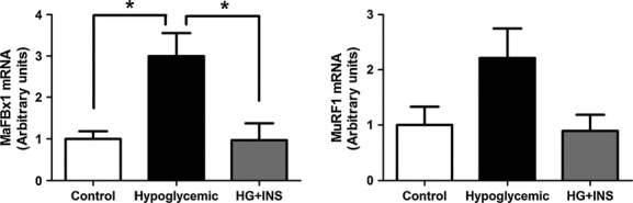
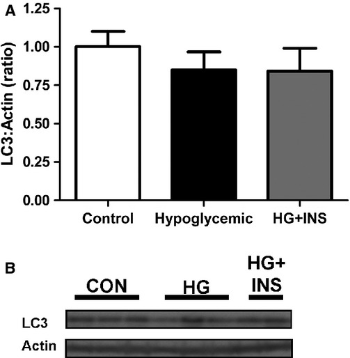
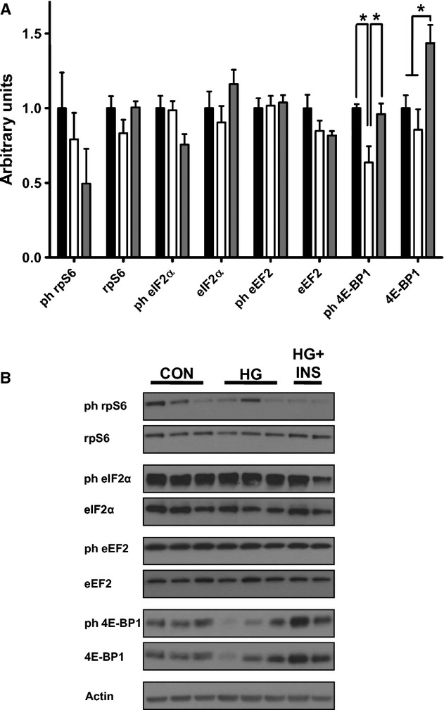
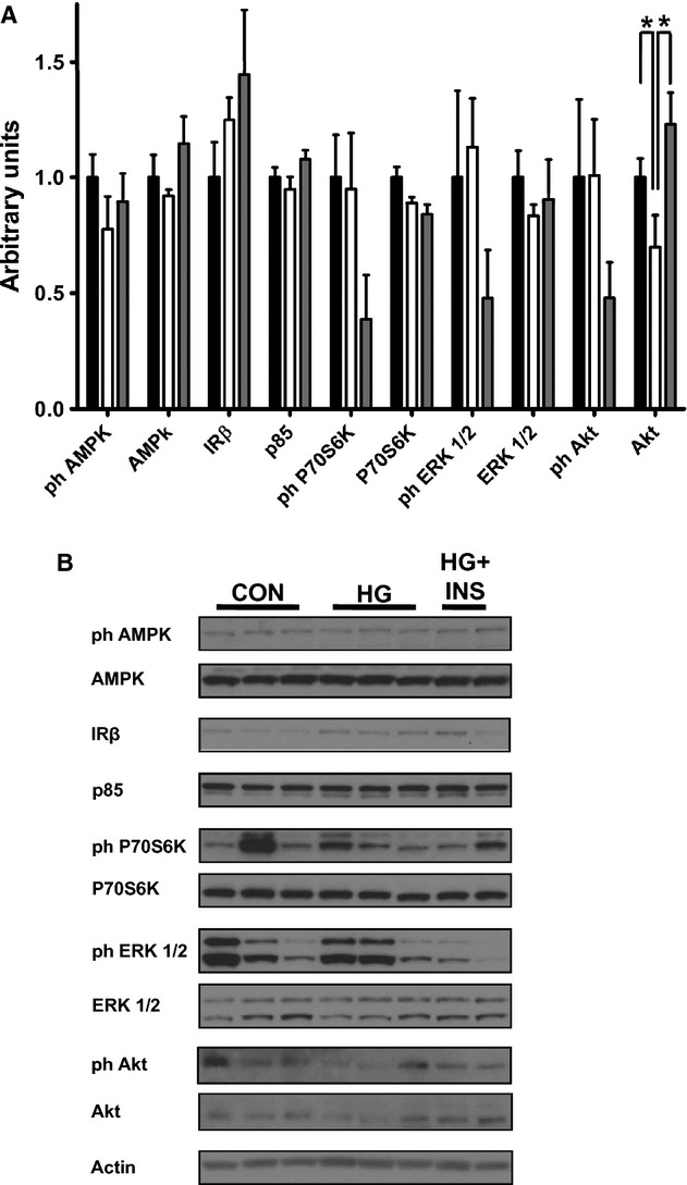
Decreased glucose transfer to the fetus is characteristic of pregnancies complicated by maternal under nutrition and placental insufficiency. Chronic experimental restriction of glucose transfer to the sheep fetus for the final 40% of gestation with a maternal insulin infusion (HG fetuses) results in fetal hypoglycemia, hypoinsulinemia, and decreased rates of fetal growth and protein accretion compared to controls (CON). Lower rates of fetal protein accretion are due to increased fetal protein breakdown and not decreased protein synthesis. However, the specific skeletal muscle pathways responsible for increased protein breakdown have not been determined. Nor has it been determined if low fetal glucose or insulin concentrations are more important for regulating these skeletal muscle protein breakdown pathways. We tested whether chronic restriction of glucose transfer to the fetus increased the ubiquitin-proteosome pathway or autophagy-lysosome pathway in fetal sheep skeletal muscle and found no evidence for an increase in the autophagy-lysosome pathway. However, HG fetuses had increase mRNA expression of MaFBx1 (twofold, P < 0.01) and a trend for increased mRNA expression of MuRF1 (P = 0.08) compared to CON. A subset of chronically hypoglycemic fetuses received an isoglycemic insulin infusion for the final 7 days of the maternal insulin infusion (HG + INS fetuses) and had MaFBx1 and MuRF1 mRNA concentrations similar to CON fetuses. These results demonstrate that fetuses exposed to sustained hypoglycemia have decreased protein accretion due to activation of the skeletal muscle ubiquitin-proteosome pathway and that a fetal hyperinsulinemic clamp can suppress this pathway even in the context of continued hypoglycemia.
Keywords: Autophagy‐Lysosome; MaFBx1; MuRF1; pregnancy; ubiquitin‐proteosome.
© 2014 The Authors. Physiological Reports published by Wiley Periodicals, Inc. on behalf of the American Physiological Society and The Physiological Society.
Figures
References
-
- Anderson M. S., Thamotharan M., Kao D., Devaskar S. U., Qiao L., Friedman J. E. 2005. Effects of acute hyperinsulinemia on insulin signal transduction and glucose transporters in ovine fetal skeletal muscle. Am. J. Physiol. Regul. Integr. Comp. Physiol.; 288:R473-R481. - PubMed
-
- Baptista I. L., Leal M. L., Artioli G. G., Aoki M. S., Fiamoncini J., Turri A. O. 2010. Leucine attenuates skeletal muscle wasting via inhibition of ubiquitin ligases. Muscle Nerve; 41:800-808. - PubMed
-
- Belkacemi L., Nelson D. M., Desai M., Ross M. G. 2010. Maternal undernutrition influences placental‐fetal development. Biol. Reprod.; 83:325-331. - PubMed
-
- Biolo G., Zhang X. J., Wolfe R. R. 1995. Role of membrane transport in interorgan amino acid flow between muscle and small intestine. Metabolism; 44:719-724. - PubMed
-
- Biolo G., Fleming R. Y., Maggi S. P., Nguyen T. T., Herndon D. N., Wolfe R. R. 2002. Inverse regulation of protein turnover and amino acid transport in skeletal muscle of hypercatabolic patients. J. Clin. Endocrinol. Metab.; 87:3378-3384. - PubMed
Grants and funding
LinkOut - more resources
Full Text Sources
Other Literature Sources
