

Correlations between skin lesions induced by anti-tumor necrosis factor-α and selected cytokines in Crohn's disease patients
- PMID: 24944497
- PMCID: PMC4051946
- DOI: 10.3748/wjg.v20.i22.7019
Correlations between skin lesions induced by anti-tumor necrosis factor-α and selected cytokines in Crohn's disease patients
Abstract
Aim: To investigate the correlation between the appearance of skin lesions and concentration of interleukin (IL)-17A, IL-23 and interferon-γ (IFN-γ) in Crohn's disease (CD) patients during anti-tumor necrosis factor-α (TNF-α) therapy
Methods: A prospective study included 30 adult patients with CD of Caucasian origin (19 men and 11 women; mean age ± SD 32.0 ± 8.6 years) during biological therapy with anti-TNF-α antibodies from January 2012 to March 2013. Eighteen patients were treated with infliximab, seven with adalimumab and five with certolizumab. Inclusion criteria were exacerbation of the underlying disease, Crohn's Disease Activity Index over 300 and the ineffectiveness of previously used non-biological therapies. Patients with a history of psoriasis, atopic dermatitis and other autoimmune skin lesions were excluded from the study. The control group consisted of 12 healthy subjects. A diagnostic survey was carried out, blood tests and careful skin examination were performed, and the serum levels of IL-17, IL-23 and IFN-γ were measured using an enzyme-linked immunosorbent assays technique. Dermatoses that have developed in the course of biological therapy in patients who had no pre-existing skin lesions of similar character were qualified as skin lesions induced by anti-TNF-α therapy.
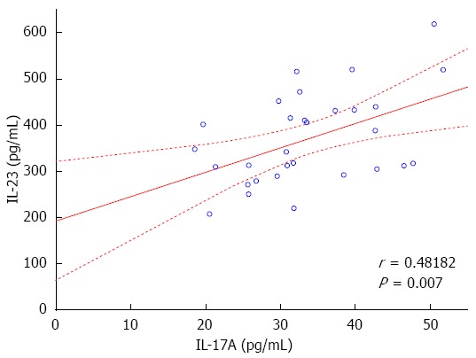
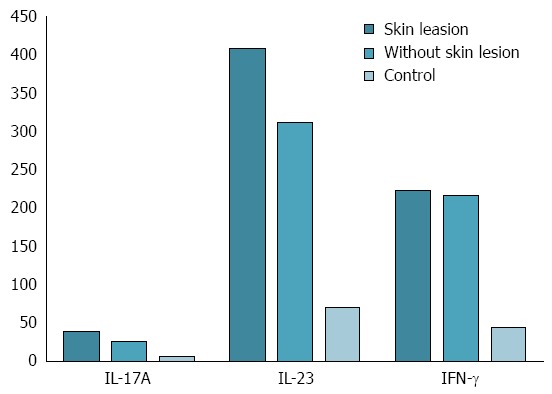
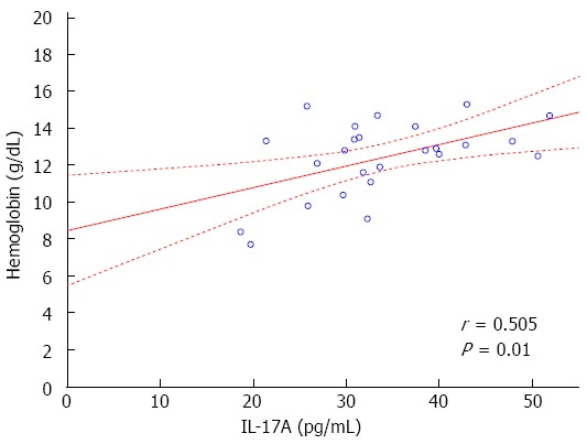
Results: Skin manifestations occurred in 18 of CD patients during the anti-TNF-α therapy (60%), in the average time of 10.16 ± 3.42 mo following the beginning of the 52-wk treatment cycle. Skin lesions observed in CD patients during biological therapy included psoriasiform lesions (44.4%), and eczema forms lesions (22.2%). In CD patients with drug induced skin lesions significantly higher levels of hemoglobin (13.3 ± 1.5 g/dL vs 10.8 ± 1.9 g/dL, P = 0.018) and hematocrit (39.9% ± 4.5% vs 34.3% ± 5.4%, P = 0.01), as well as a significantly lower level of platelets (268 ± 62 × 10(3)/μL vs 408 ± 239 × 10(3)/μL, P = 0.046) was observed compared with CD patients without skin manifestations. The concentrations of IL-17A and IL-23 in CD patients with skin lesions developed under anti-TNF-α therapy were significantly higher compared to those in patients without lesions (IL-17A: 39.01 ± 7.03 pg/mL vs 25.71 ± 4.90 pg/mL, P = 0.00004; IL-23: 408.78 ± 94.13 pg/mL vs 312.15 ± 76.24 pg/mL, P = 0.00556).
Conclusion: Skin lesions in CD patients during biological therapy may result from significantly increased concentrations of IL-17A and IL-23, which are strongly associated with TNF-α/Th1 immune pathways.
Keywords: Biological therapy; Crohn’s disease; Interferon γ; Interleukin 17A; Interleukin 23; Tumor necrosis factor-α.
Figures
References
-
- Van Assche G, Dignass A, Panes J, Beaugerie L, Karagiannis J, Allez M, Ochsenkühn T, Orchard T, Rogler G, Louis E, et al. The second European evidence-based Consensus on the diagnosis and management of Crohn’s disease: Definitions and diagnosis. J Crohns Colitis. 2010;4:7–27. - PubMed
-
- Dignass A, Van Assche G, Lindsay JO, Lémann M, Söderholm J, Colombel JF, Danese S, D’Hoore A, Gassull M, Gomollón F, et al. The second European evidence-based Consensus on the diagnosis and management of Crohn’s disease: Current management. J Crohns Colitis. 2010;4:28–62. - PubMed
-
- Ardizzone S, Puttini PS, Cassinotti A, Porro GB. Extraintestinal manifestations of inflammatory bowel disease. Dig Liver Dis. 2008;40 Suppl 2:S253–S259. - PubMed
Publication types
MeSH terms
Substances
LinkOut - more resources
Full Text Sources
Other Literature Sources
Medical
