

Imaging and pathological features of primary hepatic neuroendocrine carcinoma: An analysis of nine cases and review of the literature
- PMID: 24944650
- PMCID: PMC3961293
- DOI: 10.3892/ol.2014.1844
Imaging and pathological features of primary hepatic neuroendocrine carcinoma: An analysis of nine cases and review of the literature
Abstract
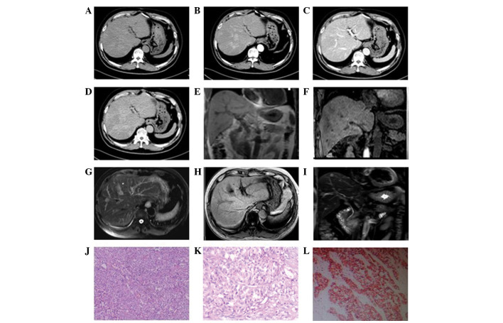
The present study aimed to analyze the imaging features and pathological basis of primary hepatic neuroendocrine carcinoma (PHNEC). A retrospective analysis of the imaging and pathological features of nine PHNEC cases was carried out at The Second Xiangya Hospital of Central South University (Changsha, China). The nine patients were subjected to dynamic contrast-enhanced computed tomography (CT) scanning of the liver and pathological diagnosis of the tissue samples. In addition, two patients were subjected to magnetic resonance imaging (MRI). CT scanning revealed the presence of single or multiple masses in the liver with a maximum diameter of 1-10 cm. These hepatic masses were of low density as showed by plain CT. These masses showed uneven or annular enhancement at their margins in the arterial phase. The venous portal phase showed consistent or declined enhancement and the delayed phase showed light enhancement in these masses. In addition, multiple intrahepatic nodules with long T1 and T2 signal intensities and obvious enhancement were observed by MRI in one patient, while intrahepatic lesions with moderate length T2 signal intensities and light enhancement not visible on the T1 image were observed in another patient. Pathological analysis revealed that the tumor cells exhibited morphological diversity. Immunohistochemical staining revealed that the tumor cells were chromogranin A- and synaptophysin-positive, and carcinoembryonic antigen-, hepatocytic antigen- and α-fetoprotein-negative. Imaging methods, including CT and MRI, are useful for the diagnosis of PHNEC; however, pathological examination is required for a final, definite diagnosis.
Keywords: computed tomography; magnetic resonance imaging; pathology; primary hepatic neuroendocrine carcinoma.
Figures


References
-
- Dogra VS, Poblete J. Metastatic carcinoid tumor in the liver. J Clin Ultrasound. 1993;21:639–641. - PubMed
-
- Yalav O, Ülkü A, Akçam TA, Demiryürek H, Doran F. Primary hepatic neuroendocrine tumor: five cases with different preoperative diagnoses. Turk J Gastroenterol. 2012;23:272–278. - PubMed
-
- Shetty PK, Baliqa SV, Balaiah K, Gnana PS. Primary hepatic neuroendocrine tumor: an unusual cystic presentation. Indian J Pathol Microbiol. 2010;53:760–762. - PubMed
-
- Ishida M, Seki K, Tatsuzawa A, et al. Primary hepatic neuroendocrine carcinoma coexisting with hepatocellular carcinoma in hepatitis C liver cirrhosis: report of a case. Surg Today. 2003;33:214–218. - PubMed
LinkOut - more resources
Full Text Sources
Other Literature Sources
Research Materials