

Pulmonary sclerosing hemangioma with lymph node metastasis: A case report and literature review
- PMID: 24944657
- PMCID: PMC3961406
- DOI: 10.3892/ol.2014.1831
Pulmonary sclerosing hemangioma with lymph node metastasis: A case report and literature review
Abstract
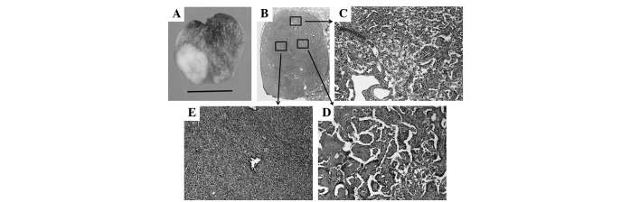
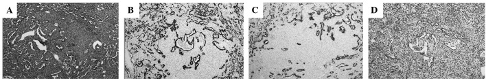
Pulmonary sclerosing hemangioma (SH) is an uncommon benign or low-grade malignant tumor. Multicentric SH and SH with lymph node metastasis have rarely been reported. The present report describes a case of pulmonary SH with lymph node metastasis in a middle-aged female. A nodule was found incidentally in the lower left lung. The patient underwent left lower pulmonary lobectomy and lymph node dissection. Histologically, the nodule demonstrated the characteristic features of SH and one of the resected lymph nodes contained a metastasis of this tumor. Thus, pulmonary SH has the potential to metastasize, a potential not suggested by histological features.
Keywords: lung; lymph node metastasis; middle-aged female; sclerosing hemangioma.
Figures


References
-
- Liebow AA, Hubbell DS. Sclerosing hemangioma (histiocytoma, xanthoma) of the lung. Cancer. 1956;9:53–75. - PubMed
-
- Devouassoux-Shisheboran M, Nicholson AG, Leslie K, Niho S. Sclerosing hemangioma. In: Travis WD, Brambilla E, Muller-Hemelink HK, Harris CC, editors. World Health Organisation Classification of Tumours: Tumors of lung, pleura, thymus and heart. IARC Press; Lyon: 2004. pp. 115–117.
-
- Keylock JB, Galvin JR, Franks TJ. Sclerosing hemangioma of the lung. Arch Pathol Lab Med. 2009;133:820–825. - PubMed
-
- Maeda R, Isowa N, Miura H, Tokuyasu H, Kawasaki Y, Yamamoto K. Bilateral multiple sclerosing hemangiomas of the lung. Gen Thorac Cariovasc Surg. 2009;57:667–670. - PubMed
LinkOut - more resources
Full Text Sources
Other Literature Sources