. 2014 Apr;7(4):1067-1072.
doi: 10.3892/ol.2014.1872. Epub 2014 Feb 11.

Clinicopathological findings in a case series of abdominopelvic solitary fibrous tumors

Affiliations

Clinicopathological findings in a case series of abdominopelvic solitary fibrous tumors

Hao Wang et al. Oncol Lett. 2014 Apr.

Abstract

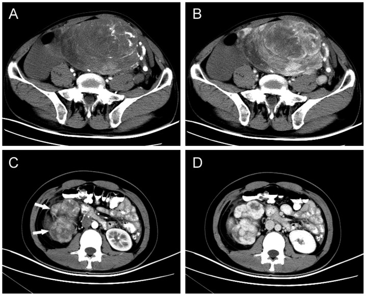
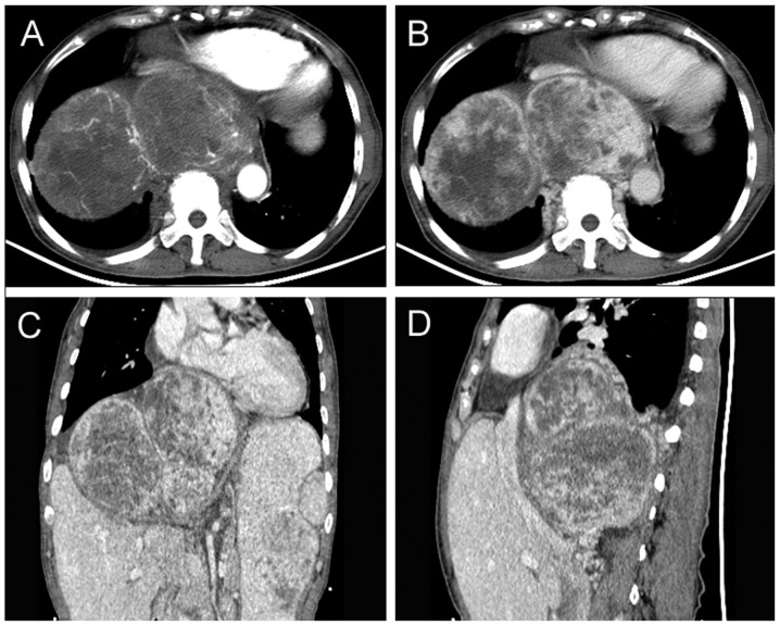
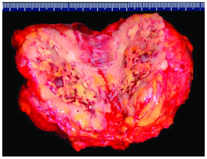
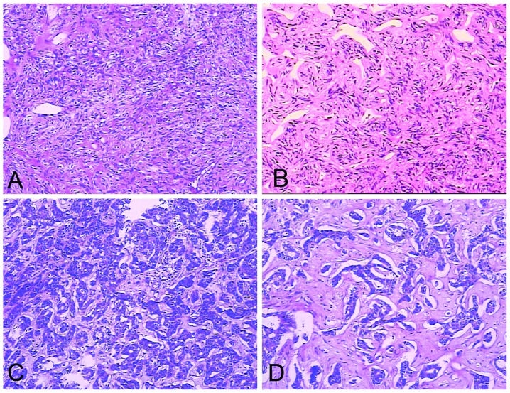
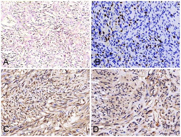
Solitary fibrous tumors (SFTs) represent a rare type of soft tissue tumor. Extrathoracic SFTs (ESFTs) in the soft tissues of the abdominopelvic cavity are extremely rare. Between January 2002 and January 2013, 10 patients were identified with abdominopelvic SFTs at the Northern Jiangsu People's Hospital. The clinicopathological data, treatment and follow-up results were retrospectively analyzed in this study. Patients included four females and six males, whose age ranged between 21 and 75 years (mean, 53.3 years). The maximum diameter of the tumors was 2.5-28 cm (mean, 12.7 cm). Two cases were diagnosed as malignant variants of ESFTs. R0 resection was performed in eight patients, while one patient underwent R1 resection, and one patient received palliative chemotherapy for an inoperable mass. Follow-up time ranged between 6 and 126 months (mean, 50 months). The patient with R1 resection suffered a local relapse, and the patient receiving palliative chemotherapy succumbed to the disease. The remaining eight patients remained free of disease. Abdominopelvic SFTs usually reveal an indolent process, although the majority of tumors in the present study were of giant size when diagnosed. The risk of local recurrence and metastasis correlates with tumor size and the histological status of surgical margins. The preferred treatment is complete resection followed by extended follow-up surveillance.

Keywords: histopathology; immunohistochemistry; solitary fibrous tumor; spindle cell tumor.


Figures

Figure 1
Figure 1
Typical imaging manifestations of abdominopelvic SFTs. (A) Arterial-phase and (B) venous-phase axial contrast-enhanced CT scan showing a well-defined, intensely enhancing mass in the pelvis with central nonenhancing areas. (C) Lobulated SFTs (indicated by arrows) of the retroperitoneum in a 21-year-old female. (D) Axial contrast-enhanced CT scan, obtained in the arterial phase, revealing a well-defined hypervascular mass with intense enhancement. SFT, solitary fibrous tumor; CT, computed tomography.
Figure 2
Figure 2
(A) Arterial-phase CT scan. The tumor shows moderate to marked enhancement, with the tumor edge visible for tortuous vascular shadow. (B) Venous-phase CT scan demonstrating sustained enhancement of the tumor. Nonenhanced areas suggest central cystic degeneration and necrosis. (C) Coronal and (D) sagittal reconstructed images: The tumor is lobulated, comprised of three masses. The heart, liver and right kidney were pressured significantly due to the tumor mass effect. CT, computed tomography.
Figure 3
Figure 3
Macroscopic appearance of an atypical malignant solitary fibrous tumor (patient no. 5). The cut surface of a well-circumscribed tumor, white- to tan-colored with deeply yellow necrotic areas.
Figure 4
Figure 4
Hematoxylin and eosin stained sections. (A) SFT consists of tightly packed round to fusiform cells with indistinct cytoplasmic borders which are arranged around a vessel. Dimensions, 10×10 cm. (B) The vessels formed a continuous, ramifying vascular network. The vessels divide and communicate with small or minute vessels which have been partly compressed and obscured by the surrounding cellular proliferation. Typically, the dividing sinusoidal vessels have an ‘antler-like’ configuration. Dimensions, 10×10 cm. (C) Malignant SFT with heightened cellularity. The tumor also demonstrates marked pleomorphism with a high level of mitotic activity (>4 mitotic fields/10 high power fields). Dimensions, 10×10 cm. (D) SFT tumor cells arranged randomly in a ‘patternless pattern’. The SFT presented marked hyalinization and had characteristic artifactual ‘cracks’ between the cells and collagen. Dimensions, 10×20 cm. SFT, solitary fibrous tumor.
Figure 5
Figure 5
Immunohistochemical staining. (A) The majority of tumors are diffusely positive for cluster of differentiation 34. Dimensions, 10×10 cm. (B) The Ki-67 labeling index is >5% in the solitary fibrous tumor identified as a malignant variant (patient no. 1). Dimensions, 10×20 cm. Tumors were positive for (C) insulin receptor in patient nos. 1, 2 and 4–8 and (D) insulin-like growth factor 1 receptor in patient nos. 1 and 4. Dimensions, 10×20 cm.

Similar articles

Cited by

References

    1. Klemperer P, Rabin CB. Primary neoplasmas of the pleura. A report of five cases. Am J Ind Med. 1992;22:4–31. - PubMed
    1. Gold JS, Antonescu CR, Hajdu C, et al. Clinicopathologic correlates of solitary fibrous tumors. Cancer. 2002;94:1057–1068. - PubMed
    1. Brunnemann RB, Ro JY, Ordonez NG, Mooney J, El-Naggar AK, Ayala AG. Extrapleural solitary fibrous tumor: a clinicopathologic study of 24 cases. Mod Pathol. 1999;12:1034–1042. - PubMed
    1. Fukunaga M, Naganuma H, Nikaido T, Harada T, Ushigome S. Extrapleural solitary fibrous tumor: a report of seven cases. Mod Pathol. 1997;10:443–450. - PubMed
    1. Hasegawa T, Matsuno Y, Shimoda T, Hasegawa F, Sano T, Hirohashi S. Extrathoracic solitary fibrous tumors: their histological variability and potentially aggressive behavior. Hum Pathol. 1999;30:1464–1473. - PubMed

LinkOut - more resources