

Two adult cases of extralobar pulmonary sequestration: A non-complicated case and a necrotic case with torsion
- PMID: 24944723
- PMCID: PMC4061149
- DOI: 10.12659/PJR.890662
Two adult cases of extralobar pulmonary sequestration: A non-complicated case and a necrotic case with torsion
Abstract
Background: This case report describes two cases of extralobar pulmonary sequestration in adults with and without torsion/necrosis.
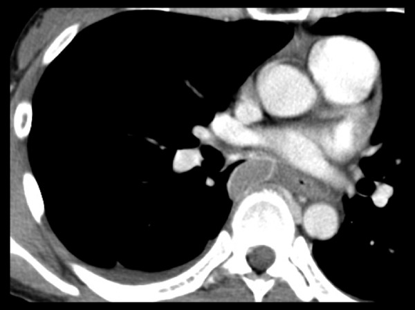
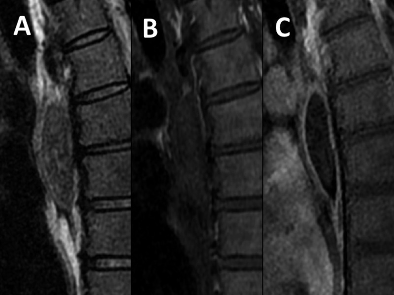
Case reports: Non-complicated extralobar pulmonary sequestration was found incidentally in a 50-year-old asymptomatic woman (Case 1), diagnosed with the presence of a branching structure in a mass lesion and blood supply from the right inferior phrenic artery. Another case of a 38-year-old woman presented with a sudden onset of back pain caused by extralobar pulmonary sequestration with torsion/necrosis (Case 2). A 4-cm fusiform mass in the paravertebral region showed enhancement in the peripheral rim only, and no feeding artery. These were the same as it had been reported typical findings in extralobar pulmonary sequestration with necrosis. On magnetic resonance imaging, the masses in both cases showed inhomogeneous low signal and branching high signal on T2-weighted images. That was characteristic for a stroma without dilated alveoli as a solid part and dilated alveoli as fluid regions.
Conclusions: By comparing those two cases, we came to a conclusion that only T2-weighted imaging reflects the native structure, even after infarction. Although differentiation from a cystic tumor with hemorrhage or infection can be problematic, inhomogeneous low signal and branching high signal on T2-weighted images may help us distinguish extralobar pulmonary sequestration from other cystic lesions.
Keywords: Bronchopulmonary Sequestration; Necrosis; Torsion, Mechanical.
Figures


References
-
- Pryce DM. Lower accessory pulmonary artery with intralobar sequestration of lung: a report of seven cases. J Path Bact. 1946;58:457–67. - PubMed
-
- Stocker JT. Sequestrations of the lung. Semin Diagn Pathol. 1986;3:106–21. - PubMed
-
- Rosado-de-Christenson ML, Frazier AA, Stocker JT, Templeton PA. Extralobar sequestration: Radiologic pathologic correlation. Radiograpics. 1993;13:425–41. - PubMed
-
- Hasleton PS. Spencer’s Pathology of the Lung. New York: McGraw-Hill; 2013. pp. 71–73.
-
- Travis WD. AFIP Atlas of nontumor Pathology, Fascicle2-Non-Neoplastic Disorders of the Lower Respiratory Tract. 2002. pp. 484–86.
Publication types
LinkOut - more resources
Full Text Sources
Other Literature Sources