Review
. 2014 Mar 19;3(4):372-83.
doi: 10.1016/j.molmet.2014.03.002. eCollection 2014 Jul.

Circadian control of glucose metabolism

Affiliations
Review

Circadian control of glucose metabolism

Andries Kalsbeek et al. Mol Metab. .

Abstract

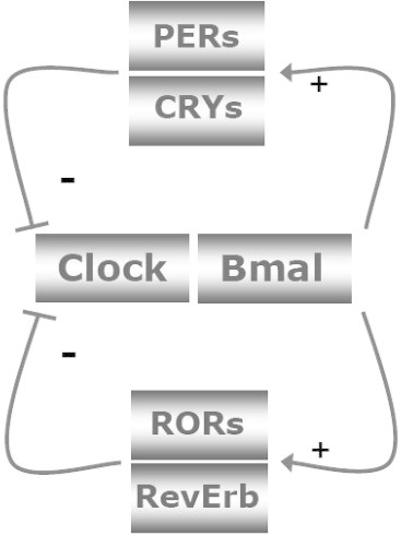
The incidence of obesity and type 2 diabetes mellitus (T2DM) has risen to epidemic proportions. The pathophysiology of T2DM is complex and involves insulin resistance, pancreatic β-cell dysfunction and visceral adiposity. It has been known for decades that a disruption of biological rhythms (which happens the most profoundly with shift work) increases the risk of developing obesity and T2DM. Recent evidence from basal studies has further sparked interest in the involvement of daily rhythms (and their disruption) in the development of obesity and T2DM. Most living organisms have molecular clocks in almost every tissue, which govern rhythmicity in many domains of physiology, such as rest/activity rhythms, feeding/fasting rhythms, and hormonal secretion. Here we present the latest research describing the specific role played by the molecular clock mechanism in the control of glucose metabolism and speculate on how disruption of these tissue clocks may lead to the disturbances in glucose homeostasis.

Keywords: Autonomic nervous system; Circadian rhythm; Diabetes; Glucose; Hypothalamus.


Figures

Figure 1
Figure 1
Simplified version of the molecular core clock mechanism. The core loop is formed by Clock:Bmal1 and Period 1–3 (Per1–3) and Cryptochrome 1 and 2 (Cry1–2). The Clock:Bmal1 heterodimer stimulates the transcription of Per1–3 and Cry1–2. Subsequently, Per's and Cry's heterodimerize, translocate to the nucleus, and inhibit Clock:Bmal1 activity. As a consequence, Clock:Bmal1 transcriptional activity drops, which reduces the transcription of Per and Cry genes, thereby activating Clock:Bmal1 again. Additional loops formed by RevErbs and RORs enhance the robustness of the core loop. Post-translational modifications of the clock proteins such as phosphorylation, ubiquitination and sumoylation greatly determine their stability and degradation, which plays a critical role in circadian cycle progression and setting the clock period. For clarity these posttranscriptional regulatory events have been left out of the figure. The feedback loops of the core clock can also regulate widely different phases of genes encoding regulatory components of energy metabolism, by binding to the appropriate promoter elements. On the other hand, changes in energy status have an impact on the molecular clock mechanism. AMPK phoshorylates Cry1 and thereby targets it for degradation. SIRT1 deacetylates, amongst others, Bmal1 and Per2, thereby decreasing the half-life of Per2.
Figure 2
Figure 2
(upper part): Plasma insulin and blood glucose responses after meal ingestion during the light period and dark period in animals on a regular feeding regimen of 6-meals-a-day. Before sampling animals had been on this regimen for 2–3 weeks. Boxes indicate meals. Despite similar meals (3.1 ± 03 g) and comparable glucose increments insulin responses significantly differ depending on the time of day. (lower part): Plasma glucose and insulin responses after the intravenous injection of a glucose bolus (500 mg/kg BW) at different times of the light/dark cycle. The maximal glucose increment at ZT14 was significantly lower than the ones at the other 5-time points. On the other hand, the total amount of insulin released did not differ between the different time points. Responses are expressed as the difference from the respective t = 0 values. Black bars indicates meals in the dark period. ZT = Zeitgeber Time; ZT12 being defined as the onset of the dark period. Adapted from Ref. (upper figure) and Ref. (lower figure).
Figure 3
Figure 3
Midsagittal view of the rat brain presenting the proposed involvement of orexin neurons in the control of the daily plasma glucose rhythm. (i) Orexin-containing neurons in the perifornical area (PF) are innervated by both glutamatergic and GABAergic projections from the biological clock (SCN). During the main part of the light period, activation of the orexin neurons by the excitatory glutamatergic inputs is prevented by the simultaneous release of the inhibitory neurotransmitter GABA (the daily activity pattern of these inputs is indicated by the lines in the yellow/blue boxes aside the projections). The circadian withdrawal of the GABAergic input at the end of the light period allows the orexin neurons to become active at the onset of darkness. (ii) Subsequently, the excitatory effect of orexin on the preganglionic neurons in the intermediolateral column (IML) of the spinal cord will (iii) activate the sympathetic input to the liver and result in increased hepatic glucose production. Orexin also stimulates glucose uptake in skeletal muscle via an action in the VMH and mediated through the sympathetic nervous system ; but, as it is not clear yet how this message is propagated from the VMH to the autonomic nervous system, this action has not been incorporated in this schema. Possibly the effect of orexin in the VMH on glucose uptake is mediated via VMH projections to the pre-autonomic neurons in the PVN. Moreover, also the effect of orexin on hepatic glucose production might in fact in involve a projection of the PF orexin neurons to the pre-autonomic neurons in the PVN, instead of, or in addition to, the direct projection of the PF orexin neurons to the spinal cord as drawn in the figure.

Similar articles

Cited by

References

    1. Peek C.B., Ramsey K.M., Marcheva B., Bass J. Nutrient sensing and the circadian clock. Trends in Endocrinology and Metabolism. 2012;23:312–318. - PMC - PubMed
    1. Stephan F.K., Zucker I. Circadian rhythms in drinking behavior and locomotor activity of rats are eliminated by hypothalamic lesions. Proceedings of the National Academy of Sciences of the United States of America. 1972;69:1583–1586. - PMC - PubMed
    1. Reppert S.M., Weaver D.R. Molecular analysis of mammalian circadian rhythms. Annual Review of Physiology. 2001;63:647–676. - PubMed
    1. Lucas R.J., Lall G.S., Allen A.E., Brown T.M. How rod, cone, and melanopsin photoreceptors come together to enlighten the mammalian circadian clock. Progress in Brain Research. 2012;199:1–18. - PubMed
    1. Balsalobre A., Damiola F., Schibler U. A serum shock induces circadian gene expression in mammalian tissue culture cells. Cell. 1998;93:929–937. - PubMed

LinkOut - more resources