Review
. 2014 Jun 2:2:702-14.
doi: 10.1016/j.redox.2014.05.006. eCollection 2014.

Mitochondrial reactive oxygen species: a double edged sword in ischemia/reperfusion vs preconditioning

Affiliations
Review

Mitochondrial reactive oxygen species: a double edged sword in ischemia/reperfusion vs preconditioning

Theodore Kalogeris et al. Redox Biol. .

Abstract

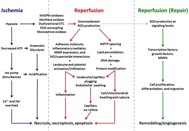
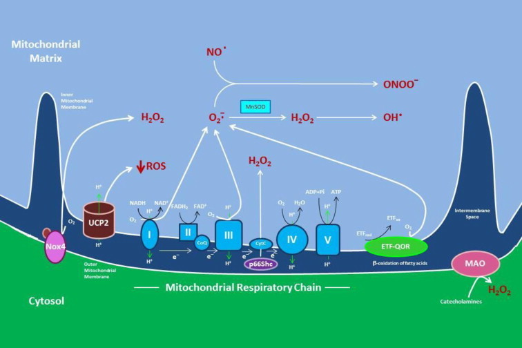
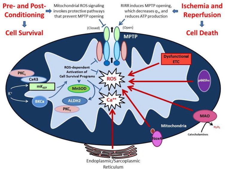
Reductions in the blood supply produce considerable injury if the duration of ischemia is prolonged. Paradoxically, restoration of perfusion to ischemic organs can exacerbate tissue damage and extend the size of an evolving infarct. Being highly metabolic organs, the heart and brain are particularly vulnerable to the deleterious effects of ischemia/reperfusion (I/R). While the pathogenetic mechanisms contributing to I/R-induced tissue injury and infarction are multifactorial, the relative importance of each contributing factor remains unclear. However, an emerging body of evidence indicates that the generation of reactive oxygen species (ROS) by mitochondria plays a critical role in damaging cellular components and initiating cell death. In this review, we summarize our current understanding of the mechanisms whereby mitochondrial ROS generation occurs in I/R and contributes to myocardial infarction and stroke. In addition, mitochondrial ROS have been shown to participate in preconditioning by several pharmacologic agents that target potassium channels (e.g., ATP-sensitive potassium (mKATP) channels or large conductance, calcium-activated potassium (mBKCa) channels) to activate cell survival programs that render tissues and organs more resistant to the deleterious effects of I/R. Finally, we review novel therapeutic approaches that selectively target mROS production to reduce postischemic tissue injury, which may prove efficacious in limiting myocardial dysfunction and infarction and abrogating neurocognitive deficits and neuronal cell death in stroke.


Figures

Fig. 1
Fig. 1
Mechanisms contributing to tissue injury in ischemia/reperfusion (I/R). Cellular hypoxia secondary to ischemia results in decreased ATP production, which in turn, disrupts ion pump function, leading to accumulation of Na+, Ca2+, and H+, with cellular acidification further promoted by a shift to anaerobic glycolysis for energy production. Activation and upregulated expression of enzymes capable of producing reactive oxygen species (ROS) and electron transport chain (ETC) dysfunction are also initiated during ischemia. These events set the stage for a burst of ROS generation when molecular oxygen is reintroduced to ischemic tissues when the blood supply is re-established. ROS-dependent expression of proinflammatory stimuli and expression of adhesion molecules by endothelial cells and leukocytes precipitates the infiltration and activation of neutrophils, T cells and monocytes. Phagocytic Nox2 activation results the respiratory burst of superoxide production that further magnifies the massive oxidative stress that directly damages virtually every biomolecule found in cells and induces the programmed cell death responses, apoptosis and necroptosis. Postischemic ROS generation also activates matrix metalloproteinases (MMPs) and other proteases that act to cleave proteins and receptors, thereby impairing their function. The net impact of these ROS-dependent events is opening of mitochondrial permeability transition pores (MPTPs), which contributes to swelling and lysis of cells. Increases in leukocyte stiffness induced by hypoxia and acidosis during ischemia lead to impaction of these cells in capillaries, an effect that is exacerbated by ROS-dependent endothelial cell swelling which in turn reduces their diameter when the blood supply is re-established. Thus, a nutritive perfusion impairment becomes prominent during reperfusion, despite repair of the precipitating ischemic event. In direct contrast to these catastrophic effects of ROS generation secondary to events occurring in ischemia and early reperfusion, oxidant production also occurs at later stages of reperfusion as tissue repair is initiated. However, ROS production occurs at lower levels that allow oxidant species to serve as signaling molecules that participate in transcriptional activation of growth factors and promote cell proliferation, differentiation and migration. The net effect of these processes is tissue and vascular remodeling, including angiogenesis. While some of these repair processes help restore organ function, others such as tissue fibrosis contribute over time to eventual organ failure. The mechanisms depicted in this figure emphasize the concept that ROS generation play key roles in all three phases of ischemia/reperfusion injury and cell death.
Fig. 2
Fig. 2
Sources of reactive oxygen species (ROS) in mitochondria. The activity of the electron transport chain generates a relatively small flux of ROS under normal conditions, but its production can be greatly magnified by events occurring during ischemia and reperfusion. Complex I (NADH dehydrogenase) and complex III (coenzyme Q (CoQ) and cytochrome C oxidoreductase) produce superoxide (O2-), which leads to hydrogen peroxide (H2O2) formation by spontaneous dismutation or via the enzymatic action of manganese superoxide dismutase (MnSOD). In the presence of transition metals, H2O2 can form the highly reactive hydroxyl radical (OH) superoxide can also interact with nitric oxide (NO) to form reactive nitrogen oxide species such as peroxynitrite (ONOO-), which produce cellular dysfunction by S-nitrosylating proteins. ROS generated by complex I are released into the mitochondrial matrix, while superoxide produced by complex III can occur in both the mitochondrial matrix and the intermembrane space between the outer and inner mitochondrial membranes. Other sources of mitochondrial superoxide are enzymes glycerol-3-phosphate dehydrogenase (G3PD), the growth factor adaptor p66Shc, and NADPH oxidase-4 (Nox4). ß-oxidation of fatty acids can also result in mitochondrial superoxide generation secondary to oxidation of electron transferring protein (ETF) by the catalytic activity of the electron transferring flavoprotein ubiquinone oxidoreductase (ETF-QOR), another enzyme expressed on the mitochondrial inner membrane. Monoamine oxidase (MAO), which is localized to the outer mitochondrial membrane, catalyzes the formation of H2O2 secondary to catecholamine metabolism. Not depicted are the mitochondrial enzymes aconitase and dihydroorotate, which can produce superoxide, but their role in ischemia/reperfusion is uncertain.The mitochondrial anion carrier, uncoupling protein-2 (UCP2), functions to separate oxidative phosphorylation from ATP synthesis with energy dissipated as heat, a phenomenon referred to as the mitochondrial proton leak. UCP2 acts to facilitate the transfer of anions from the inner to the outer mitochondrial membrane and the return transfer of protons from the outer to the inner mitochondrial membrane. They also reduce the mitochondrial membrane potential in mammalian cells. Although it was originally thought to play a role in nonshivering thermogenesis, obesity, diabetes and atherosclerosis, it now appears that the main function of UCP2 is the control of mitochondria-derived ROS.
Fig. 3
Fig. 3
Generation of reactive oxygen species (ROS) by mitochondria (mitoROS) is a nexus for both activation of cell survival programs that mediate the effect of conditioning stimuli to enhance tolerance to ischemia/reperfusion (I/R) and serves as a focal point for overexuberant ROS-induced ROS release that contributes to the pathogenesis of cell injury in I/R. On the one hand, ROS triggers the activation of cell survival programs in responses to a number of mildly noxious stimuli such as short bouts of ischemia or antecedent ethanol exposure or pharmacologic agents (ethanol or activators of mitochondrial ATP-sensitive potassium (mKATP) or large conductance, calcium-activated potassium (BKCa) channels). The enhanced tolerance to ischemia invoked by these mitoROS-dependent conditioning stimuli, which can be delivered before (preconditioning), during (preconditioning) or at the onset of reperfusion (postconditioning), activate protective protein kinases such as PKCe, the expression of prosurvival genes (e.g., heme oxygenase-1) and mitochondrial antioxidant defenses (e.g., MnSOD, aldehyde dehydrogenase-1 or ALDH2), as well as targeting the mitochondrial permeability transition pore (MPTP) to maintain the channel in a closed state. On the other hand, overexuberant ROS generation at the onset of reperfusion, driven by ROS-induced ROS release that is fueled by electron transport chain dysfunction, especially at complexes I and III, and enhanced activities of p66Shc, monoamine oxidase (MAO), and NADPH oxidase-4 (Nox4) in mitochondria, causes the MPTP to open, leading to swelling, cell disruption and death. Not depicted is the effect of oxidants to alter the balance of mitochondrial fission and fusion in conditioning and I/R, which emerging evidence has implicated as contributory to both processes. The dual nature of ROS as protective vs damaging species relates to the type of ROS generated in particular circumstances, their concentration, and/or compartmental localization of their production.

References

    1. Andreadou I., Iliodromitis E.K., Farmakis D., Kremastinos D.T. To prevent, protect and save the ischemic heart: antioxidants revisited. Expert Opinion on Therapeutic Targets. 2009;13:945–956. 19534573 - PubMed
    1. Bath P.M., Gray L.J., Bath A.J., Buchan A., Miyata T., Green A.R. Effects of NXY-059 in experimental stroke: an individual animal meta-analysis. British Journal of Pharmacology. 2009;157:1157–1171. 19422398 - PMC - PubMed
    1. Jaxa-Chamiec T., Bednarz B., Herbaczynska-Cedro K., Maciejewski P., Ceremuzynski L. Effects of vitamins C and E on the outcome after acute myocardial infarction in diabetics: aretrospective, hypothesis-generating analysis from the MIVIT study. Cardiology. 2009;112:219–223. 18698138 - PubMed
    1. Vivekananthan D.P., Penn M.S., Sapp S.K., Hsu A., Topol E.J. Use of antioxidant vitamins for the prevention of cardiovascular disease: meta-analysis of randomised trials. Lancet. 2003;361:2017–2023. 12814711 - PubMed
    1. Kalogeris T., Baines C.P., Krenz M., Korthuis R.J. Cell biology of ischemia/reperfusion injury. International Review of Cell and Molecular Biology. 2012;298:229–317. 22878108 - PMC - PubMed

Publication types

MeSH terms