. 2014 Jun 19;9(6):e100637.
doi: 10.1371/journal.pone.0100637. eCollection 2014.

Expression of multiple transgenes from a single construct using viral 2A peptides in Drosophila

Affiliations

Expression of multiple transgenes from a single construct using viral 2A peptides in Drosophila

Richard W Daniels et al. PLoS One. .

Abstract

Expression of multiple reporter or effector transgenes in the same cell from a single construct is increasingly necessary in various experimental paradigms. The discovery of short, virus-derived peptide sequences that mediate a ribosome-skipping event enables generation of multiple separate peptide products from one mRNA. Here we describe methods and vectors to facilitate easy production of polycistronic-like sequences utilizing these 2A peptides tailored for expression in Drosophila both in vitro and in vivo. We tested the separation efficiency of different viral 2A peptides in cultured Drosophila cells and in vivo and found that the 2A peptides from porcine teschovirus-1 (P2A) and Thosea asigna virus (T2A) worked best. To demonstrate the utility of this approach, we used the P2A peptide to co-express the red fluorescent protein tdTomato and the genetically-encoded calcium indicator GCaMP5G in larval motorneurons. This technique enabled ratiometric calcium imaging with motion correction allowing us to record synaptic activity at the neuromuscular junction in an intact larval preparation through the cuticle. The tools presented here should greatly facilitate the generation of 2A peptide-mediated expression of multiple transgenes in Drosophila.


Conflict of interest statement

Competing Interests: The authors have declared that no competing interests exist.

Figures

Figure 1
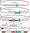
Figure 1. Outline of vector design for cloning 2A peptide-linked transgenes.
A. Diagram of pC5-Kan 2A, a vector containing a codon-optimized 2A peptide coding sequence flanked on each side by multiple cloning sites that facilitates cloning. Restriction sites in frame with the 2A sequence are shown in red. B. General procedure for making multimeric 2A-linked transgenes. A fragment cut at the 5′ end with BamHI and at the 3′ end with any of the unique restriction enzymes following the 2A coding sequence (boxed region) can then be inserted 3′ of the 2A sequence in another vector cut with BglII and the second chosen enzyme, creating the vector shown in panel C. This process creates a BamHI/BglII hybrid site that is in frame with the first coding sequence (red arrow), and leaves the BamHI and BglII sites unique so they can be used again if required. The process of adding a gene-2A cassette can be repeated using this method to create the desired construct. Examples are shown at the bottom. D. Diagram of an expression vector demonstrating the final step of the cloning procedure. The pUAS-C5 vector contains the same multiple cloning site as the pC5-Kan shuttle vector and has been modified with an attB site for phiC31-mediated integration into genomic attP sites.
Figure 2
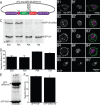
Figure 2. Demonstration of separation of proteins linked by different 2A peptides in cultured cells.
A. Diagram of vector used to transform S2 cells to express myristylated GFP and nuclear-localized dsRed. B. Single optical sections from confocal micrographs of fixed S2 cells transfected with vector shown in A. Native GFP and dsRed fluorescence are shown in the leftmost and center panels, respectively. The rightmost panel shows the merged image. While most GFP and dsRed fluorescence is localized correctly, some perinuclear colocalization can be seen. In the absence of a 2A peptide (panel B5), all GFP and dsRed signals colocalize and are enriched in a ring surrounding the nucleus. Similar perinuclear puncta observed in merged image panels (B1’’-B4’’) likely indicate full-length (unseparated) myrGFP-2A-dsRednls protein. C. Example western blot from transfected S2 cells using an anti-GFP antibody demonstrating separation efficiency of different 2A peptides. The top band runs at approximately 75 kDa and is unseparated myrGFP-2A-dsRednls, while the bottom band runs at approximately 37 kDa and is separated myrGFP-2A (GFP-2A*) only. Slight differences in running speed are due to differences in 2A peptide length. D. Histogram showing separation efficiency of 2A peptides tested in S2 cells. Error bars indicate SEM, n = 5. The P2A and T2A peptides yield the most complete separation (one-way ANOVA with Bonferonni post-test; p<0.001 vs. P2A and T2A indicated by asterisks). E. Western blot showing ratiometric expression of proteins before and after 2A peptide. The lower band is GFP-2A and the upper band is GFP-dsRed-nls. F. The intensity of each band was quantified and plotted in the histogram. The average normalized intensity of the lower band is 98+/−2 versus 93+/−7 for the upper band. Error bars indicate SEM, n = 2.
Figure 3
Figure 3. Testing the separation efficiency of 2A peptides in vivo.
A. Diagram of the DNA construct injected into flies placing expression of myrGFP-2A-dsRed-nls under the control of Gal4. B. Single confocal sections showing separation of myrGFP and dsRed-nls in salivary gland cells from third instar larvae in which UAS-myrGFP-2A-dsRednls is driven by act5c-Gal4. The first panel for each peptide version shows DNA (stained with ToPro3), the second panel shows dsRed-nls fluorescence, the third shows GFP fluorescence, and the last panel a merged image of all three channels. C. Volume maximum intensity projection of confocal micrographs from larval muscle cells expressing myrGFP-2A-dsRed-nls driven by how24B–Gal4. DsRed and GFP fluorescence are shown in the first two panels, respectively, and the third panel shows the merged image. Unseparated protein products appears as aggregates. D. Analysis of dsRed/GFP aggregates in muscle cells. More and larger aggregates are detected in cells expressing E2A- and F2A-linked transgenes, N = 2 cells per genotype. E. Western blot from adult fly tissue expressing myrGFP-2A-dsRed-nls driven by tubulin-Gal4 or LexAop-myrGFP driven by act5c-LexA (last lane) as a negative control. The upper band representing unseparated protein product cannot be seen at this exposure (asterisk is next to non-specific band also seen when myrGFP is expressed alone), despite prevalence of lower band representing separated GFP-2A* protein.
Figure 4
Figure 4. Application of the P2A peptide system for ratiometric calcium imaging at the larval NMJ.
A. Dual-channel imaging of tdTomato (top) and GCaMP5G (bottom) at the neuromuscular junctions on muscle 13 of a dissected third instar larva expressing UAS-tdTomato-P2A–GCaMP5G driven by n-syb-Gal4. B. Fluorescence records from MN13-Ib synaptic boutons responding to electrical stimulation of the segmental nerve at the indicated frequencies. C. GCaMP5G fluorescence data from B normalized to the simultaneously collected tdTomato signal. D. Histogram showing peak normalized GCaMP5G fluorescence intensity as a function of stimulation frequency (n = 6 larvae, mean +/− SEM). E. Dual-channel imaging of tdTomato (top) and GCaMP5G (bottom) at the neuromuscular junctions on muscle 13 taken through the cuticle of an intact larva. F. Fluorescence records from synaptic boutons responding to endogenous bursts of action potential firing. Note the large fluctuations in tdTomato signal that are indicative of muscle contraction and associated movement artifacts. G. GCaMP5G fluorescence from panel F normalized to tdTomato signal, which reveals a more biologically-relevant signal.

References

    1. Kaufman RJ, Davies MV, Wasley LC, Michnick D (1991) Improved vectors for stable expression of foreign genes in mammalian cells by use of the untranslated leader sequence from EMC virus. Nucleic Acids Res 19: 4485–4490. - PMC - PubMed
    1. Ryan MD, King AM, Thomas GP (1991) Cleavage of foot-and-mouth disease virus polyprotein is mediated by residues located within a 19 amino acid sequence. J Gen Virol 72 (Pt 11): 2727–2732. - PubMed
    1. Ryan MD, Drew J (1994) Foot-and-mouth disease virus 2A oligopeptide mediated cleavage of an artificial polyprotein. EMBO J 13: 928–933. - PMC - PubMed
    1. Donnelly ML, Hughes LE, Luke G, Mendoza H, ten Dam E, et al. (2001) The 'cleavage' activities of foot-and-mouth disease virus 2A site-directed mutants and naturally occurring '2A-like' sequences. J Gen Virol 82: 1027–1041. - PubMed
    1. Szymczak AL, Workman CJ, Wang Y, Vignali KM, Dilioglou S, et al. (2004) Correction of multi-gene deficiency in vivo using a single 'self-cleaving' 2A peptide-based retroviral vector. Nat Biotechnol 22: 589–594. - PubMed

Publication types

MeSH terms