

Oxidative damage and autophagy in the human trabecular meshwork as related with ageing
- PMID: 24945152
- PMCID: PMC4063984
- DOI: 10.1371/journal.pone.0098106
Oxidative damage and autophagy in the human trabecular meshwork as related with ageing
Abstract
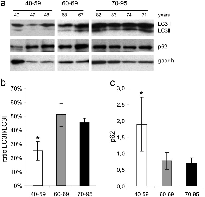
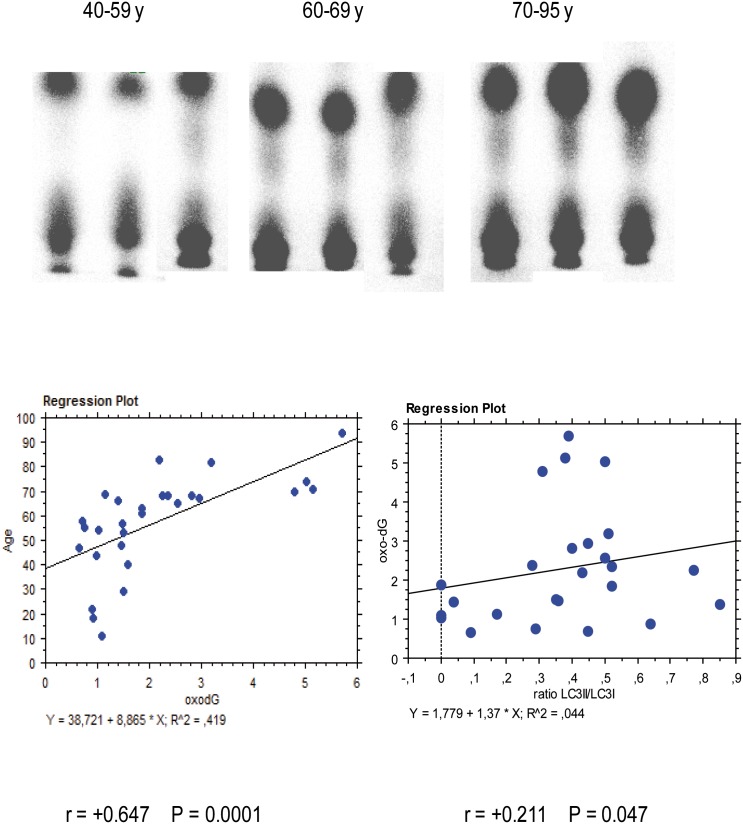
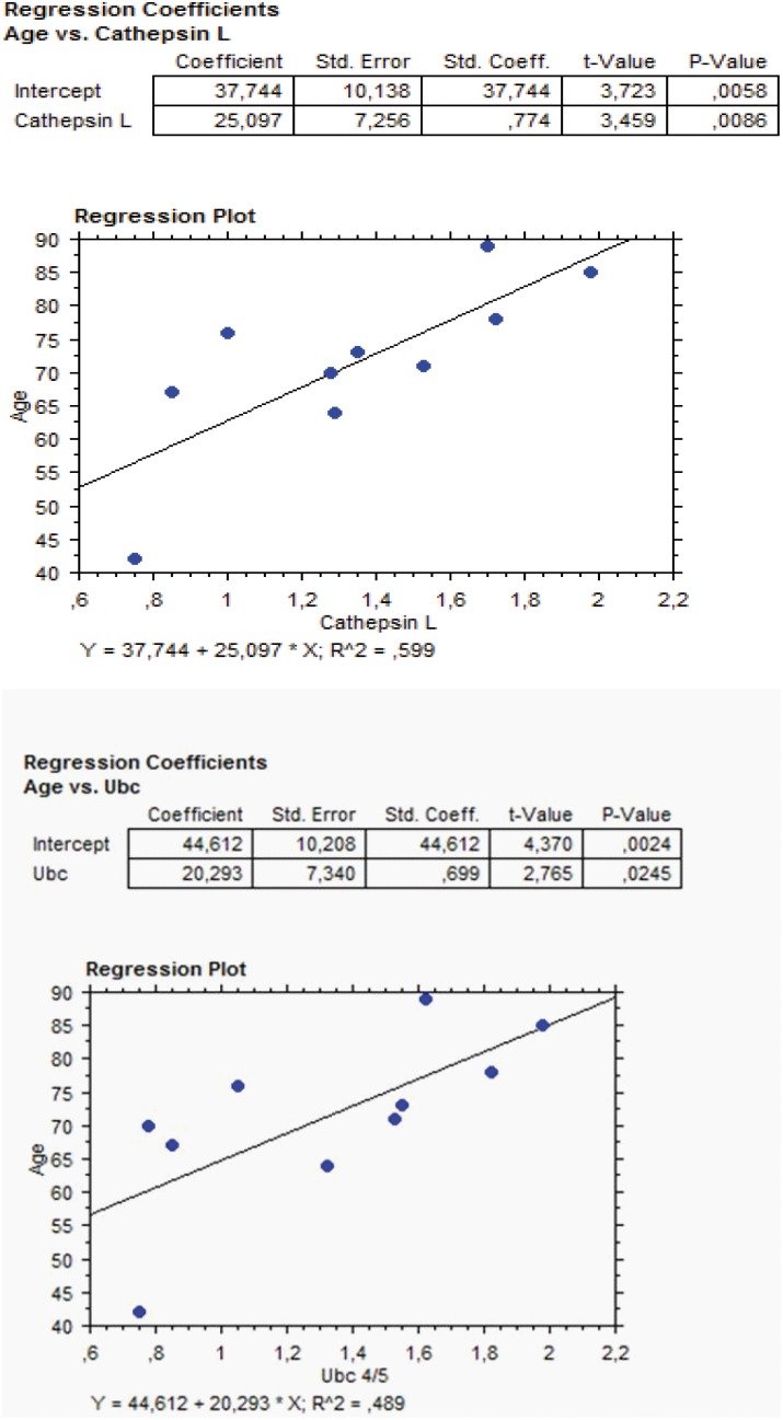
Autophagy is an intracellular lysosomal degradation process induced under stress conditions. Autophagy also plays a major role in ocular patho-physiology. Molecular aging does occur in the trabecular meshwork, the main regulator of aqueous humor outflow, and trabecular meshwork senescence is accompanied by increased oxidative stress. However, the role of autophagy in trabecular meshwork patho-physiology has not yet been examined in vivo in human ocular tissues. The purpose of the herein presented study is to evaluate autophagy occurrence in ex-vivo collected human trabecular meshwork specimens and to evaluate the relationship between autophagy, oxidative stress, and aging in this tissue. Fresh trabecular meshwork specimens were collected from 28 healthy corneal donors devoid of ocular pathologies and oxidative DNA damage, and LC3 and p62 protein expression analyzed. In a subset of 10 subjects, further to trabecular meshwork proteins, the amounts of cathepesin L and ubiquitin was analyzed by antibody microarray in aqueous humor. Obtained results demonstrate that autophagy activation, measured by LC3II/I ratio, is related with. oxidative damage occurrence during aging in human trabecular meshwork. The expression of autophagy marker p62 was lower in subjects older than 60 years as compared to younger subjects. These findings reflect the occurrence of an agedependent increase in the autophagy as occurring in the trabecular meshwork. Furthermore, we showed that aging promotes trabecular-meshwork senescence due to increased oxidative stress paralleled by autophagy increase. Indeed, both oxidative DNA damage and autophagy were more abundant in subjects older than 60 years. These findings shed new light on the role of oxidative damage and autophagy during trabecular-meshwork aging.
Conflict of interest statement
Figures
References
-
- Cuervo AM, Bergamini E, Brunk UT, Dröge W, French M, et al. (2005) Autophagy and aging: the importance of maintaining “clean” cells. Autophagy 1: 131–140. - PubMed
-
- Mizushima N (2007) Autophagy: process and function. Genes Dev 21: 2861–2873. - PubMed
-
- Tasdemir E, Chiara Maiuri M, Morselli E, Criollo A, D’Amelio M, et al. (2008) A dual role of p53 in the control of autophagy. Autophagy 4: 810–814. - PubMed
-
- Rubinsztein DC, Gestwicki JE, Murphy LO, Klionsky DJ (2007) Potential therapeutic applications of autophagy. Nat Rev Drug Discov 6: 304–312. - PubMed
Publication types
MeSH terms
Substances
Grants and funding
LinkOut - more resources
Full Text Sources
Other Literature Sources
Medical
