. 2014 Jun 19;8(6):e2949.
doi: 10.1371/journal.pntd.0002949. eCollection 2014 Jun.

Regulation of Schistosoma mansoni development and reproduction by the mitogen-activated protein kinase signaling pathway

Affiliations

Regulation of Schistosoma mansoni development and reproduction by the mitogen-activated protein kinase signaling pathway

Luiza Freire de Andrade et al. PLoS Negl Trop Dis. .

Erratum in

  • PLoS Negl Trop Dis. 2014 Jul;8(7):e3081

Abstract

Background: Protein kinases are proven targets for drug development with an increasing number of eukaryotic Protein Kinase (ePK) inhibitors now approved as drugs. Mitogen-activated protein kinase (MAPK) family members connect cell-surface receptors to regulatory targets within cells and influence a number of tissue-specific biological activities such as cell proliferation, differentiation and survival. However, the contributions of members of the MAPK pathway to schistosome development and survival are unclear.

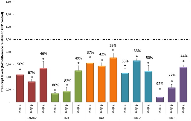
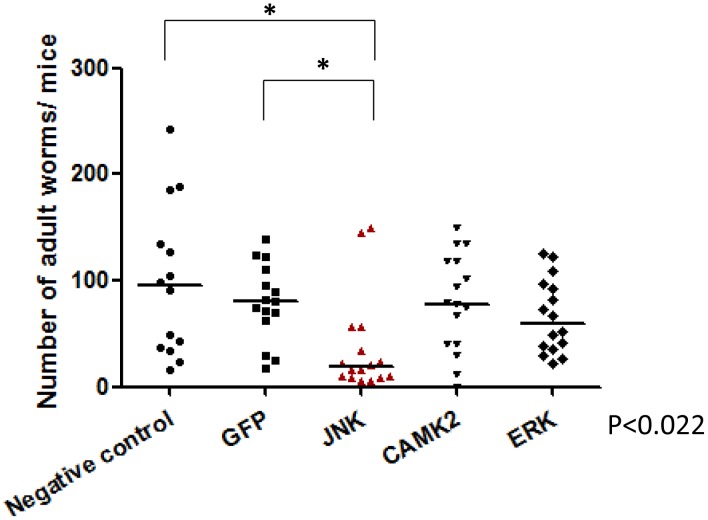
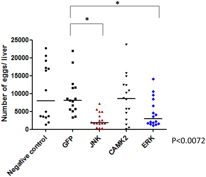
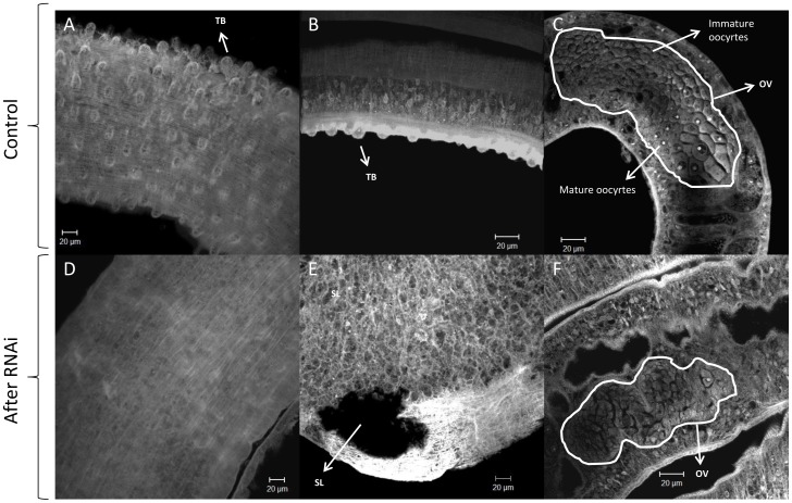
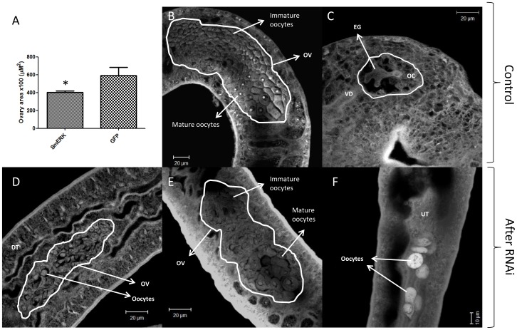
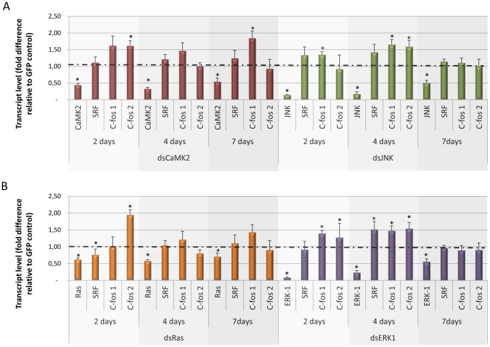
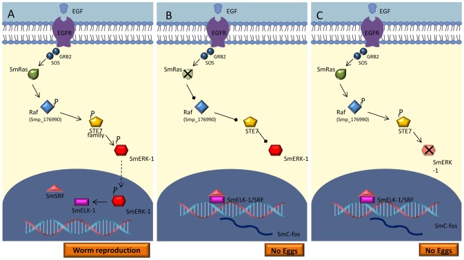
Methodology/principal findings: We employed RNA interference (RNAi) to elucidate the functional roles of five S. mansoni genes (SmCaMK2, SmJNK, SmERK1, SmERK2 and SmRas) involved in MAPK signaling pathway. Mice were injected with post-infective larvae (schistosomula) subsequent to RNAi and the development of adult worms observed. The data demonstrate that SmJNK participates in parasite maturation and survival of the parasites, whereas SmERK are involved in egg production as infected mice had significantly lower egg burdens with female worms presenting underdeveloped ovaries. Furthermore, it was shown that the c-fos transcription factor was overexpressed in parasites submitted to RNAi of SmERK1, SmJNK and SmCaMK2 indicating its putative involvement in gene regulation in this parasite's MAPK signaling cascade.

Conclusions: We conclude that MAPKs proteins play important roles in the parasite in vivo survival, being essential for normal development and successful survival and reproduction of the schistosome parasite. Moreover SmERK and SmJNK are potential targets for drug development.


Conflict of interest statement

The authors have declared that no competing interests exist.

Figures

Figure 1
Figure 1. Evolutionary relationships of ERK and JNK proteins.
Evolutionary relationships of 34 ERK and JNK proteins encoded by schistosome parasites (S. haematobium, S. japonicum and S. mansoni [different shades of blue]), Caenorhabditis elegans (green), Drosophila melanogaster (yellow), and Homo sapiens (red) as inferred by Bayesian analysis. Experimentally characterized proteins are indicated by an Erlenmeyer symbol. Different background colors highlight two clades: one containing ERK proteins and another containing JNK proteins. Support values were computed by posterior probability. The analysis was performed with conserved amino acid sequences corresponding to the catalytic domain (PF00069). Mixed models were selected as implemented in MrBayes with 10 million generations sampled every 100 generations.
Figure 2
Figure 2. Transcript levels of target genes in schistosomula 2, 4, and 7 days after exposure to dsRNA.
Bar graph indicating the relative steady-state transcript levels of SmCaMK2 (red), SmJNK (green), SmRas (orange), SmERK-2 (blue), and SmERK1 (purple) genes after 2, 4, and 7 days after dsRNA exposure. For each dsRNA tested, data are represented as mean fold-differences (+/−SE) relative to GFP control (1.00 – dashed line). Transcript levels were determined by qPCR and data analyzed using the ΔΔCt method , followed by statistical analysis using the Mann-Whitney U-test. Data were generated from 3 independent experiments, each one in duplicate, and all the data shown is statistically different from GFP controls.
Figure 3
Figure 3. Survival of the parasite after RNAi of MAPKs in vitro and subsequent transfer into mice.
Schistosomula were treated with GFP, SmJNK, SmCaMK2, and SmERK1 dsRNAs for two days and then injected into mice. After 37 days adult worms were recovered and counted. Each symbol in the chart represents worm counts from one mouse and the horizontal lines are median values per treatment group. Data were generated from 3 independent experiments and all treatments were statistical analyzed using Mann-Whitney U-test within each experiment (P≤0.05). The asterisk indicates a significance value of P<0.022 for RNAi of SmJNK relative to the GFP control.
Figure 4
Figure 4. Hepatic egg counts after RNAi of MAPKs in vitro and subsequent transfer of parasites into mice.
Schistosomula were treated with GFP, SmJNK, SmCaMK2 and SmERK1 dsRNAs for two days in vitro and then injected into mice. After 37 days of parasite eggs per liver digest were counted. Each symbol in the chart represents worm counts from one mouse and the horizontal lines are median values per treatment group. Data were generated from 3 independent experiments and all treatments were statistical analyzed using Mann-Whitney U-test within each experiment (P≤0.05). The asterisk indicates a significance value of SmJNK and SmERK P<0.0072 relative to the GFP control.
Figure 5
Figure 5. Morphology of adult worms after RNAi of SmJNK in vitro and subsequent transfer of parasites into mice.
Adult 37-day-old worms were fixed and stained, and visualized by confocal microscopy as described in the text. A, B and C show normal worms treated with GFP dsRNA, whereas D, E and F show morphological changes in worms treated with SmJNK dsRNA. A and B - the tubercules (TB) are highlighted on the tegument; C – female worm ovary (OV) showing immature and mature oocytes; D – muscular structure of a worm without tubercules; E- subtegumentar lesion (SL); F- immature ovary.
Figure 6
Figure 6. Morphology of adult female worms after RNAi of SmERK1/2 in vitro and subsequent transfer of parasites into mice.
Adult 37-day-old worms were fixed and stained, and visualized by confocal microscopy as described in the text. A - mean of females' ovary area (µM2) of SmERK-knockdown and control showing a significant size reduction; B and C show normal worms treated with GFP dsRNA where the ovary (OV) with immature and mature oocytes, an egg (EG) and the vitelloduct (VD) are visible; D, E and F show morphological changes in worms treated with SmERK dsRNA where the ovary (D) present no mature oocytes (D) or even when mature oocytes are visible (E) an unexpected phenotype (a lot of oocytes) is observed in the uterus (UT) (F). The eggs shoud be fully formed in the uterus as showing in (C). Statistical analyses were performed using Mann-Whitney U-test, P≤0.05; n = 5).
Figure 7
Figure 7. Transcript levels of SmSRF and Smc-fos1 and Smc-fos2 genes 2, 4, and 7 days after schistosomula were exposed to various MAPK dsRNAs.
Bar graph indicating the relative steady-state transcript levels of (A) SmCaMK2 (red), SmJNK (green) and (B) SmRas (orange), SmERK1(purple) genes after 2, 4, and 7 days of dsRNA exposure. For each dsRNA tested, data are represented as mean fold-differences (+/−SE) relative to GFP control (1.00). Transcript levels were determined by qPCR and data analyzed using the ΔΔCt method followed by statistical analysis using the Mann-Whitney U-test. Data were generated from 3 independent experiments, each one in duplicate. Significance levels (*) were set at P≤0.05.
Figure 8
Figure 8. Hypothetical S. mansoni MAPK signaling pathway.
(A) EGF activates the Ras/ERK signaling pathway. EGFR transmits the signal to the intracellular environment through the activation of Ras and sequential phosphorylation of SmRAF (ePK of TKL group and raf family), SmSTE7 (ePK of STE group and STE7family) and SmERK (ePK of CMGC group, MAPK family and ERK1/2 subfamily). Activated ERK translocates to the nucleus and inhibits the formation of the elk1/SRF complex and, in this case, oviposition remains constant. In B and C, SmRas or SmERK activity is interfered with (via RNAi) and the signal is not transmitted. Elk1 forms a complex with SRF which binds to the c-Fos promoter and this initiates c-Fos transcription that subsequently prevents the egg laying.

References

    1. Allen JE, Maizels RM (2011) Diversity and dialogue in immunity to helminths. Nature Reviews Immunology 11: 375–388. - PubMed
    1. Pearce EJ, Sher A (1987) Mechanisms of immune evasion in schistosomiasis. Contrib Microbiol Immunol 8: 219–232. - PubMed
    1. Han ZG, Brindley PJ, Wang SY, Chen Z (2009) Schistosoma genomics: new perspectives on schistosome biology and host-parasite interaction. Annu.Rev.Genomics Hum.Genet 10: 211–240. - PubMed
    1. Wilkinson MG, Millar JB (2000) Control of the eukaryotic cell cycle by MAP kinase signaling pathways. FASEB J 14: 2147–2157. - PubMed
    1. Hazzalin CA, Mahadevan LC (2002) MAPK-regulated transcription: A continuously variable gene switch? Nature Reviews Molecular Cell Biology 3: 30–40. - PubMed

Publication types

Substances

LinkOut - more resources