. 2014 Jun 19;10(6):e1004415.
doi: 10.1371/journal.pgen.1004415. eCollection 2014 Jun.

CPF-associated phosphatase activity opposes condensin-mediated chromosome condensation

Affiliations

CPF-associated phosphatase activity opposes condensin-mediated chromosome condensation

Vincent Vanoosthuyse et al. PLoS Genet. .

Abstract

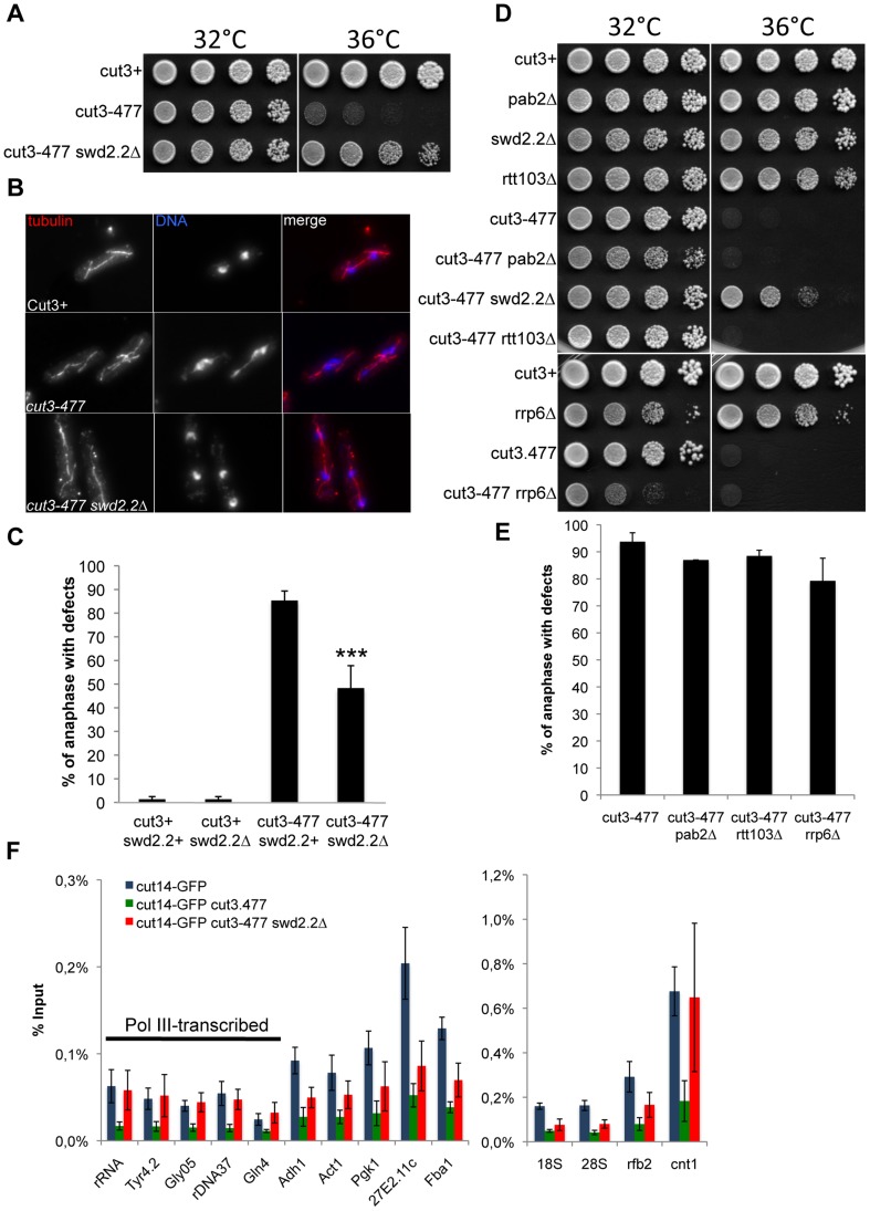
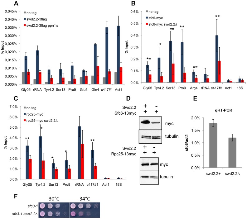
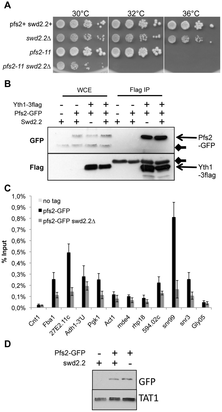
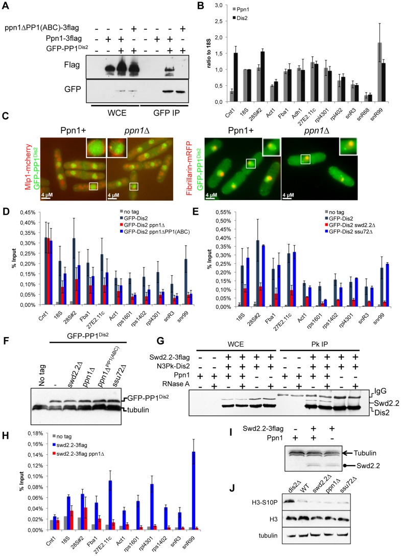
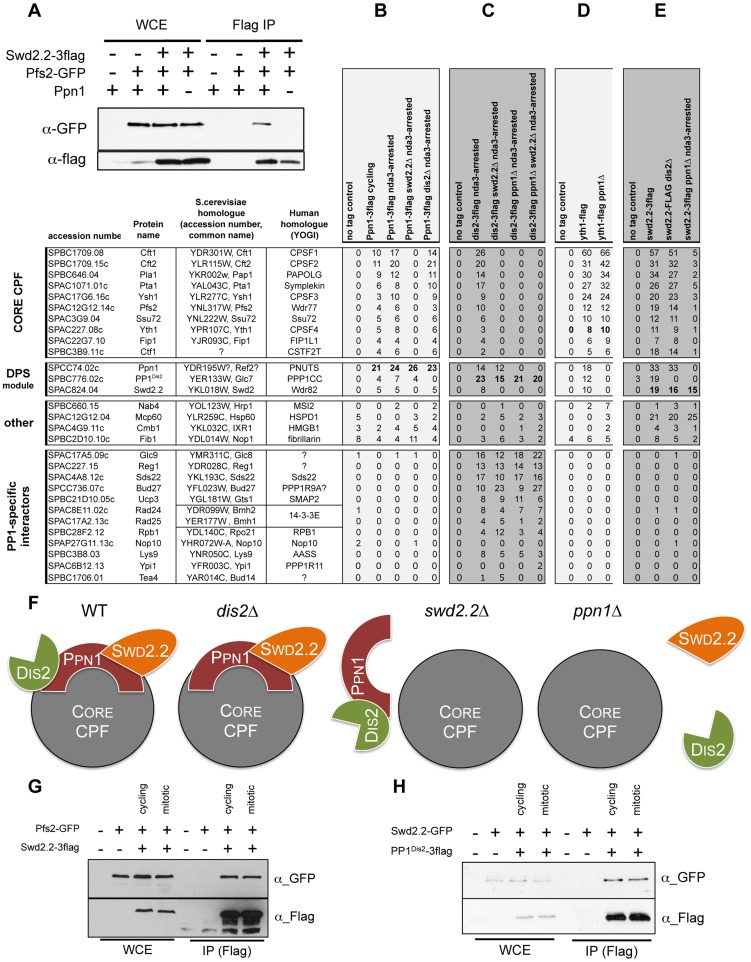
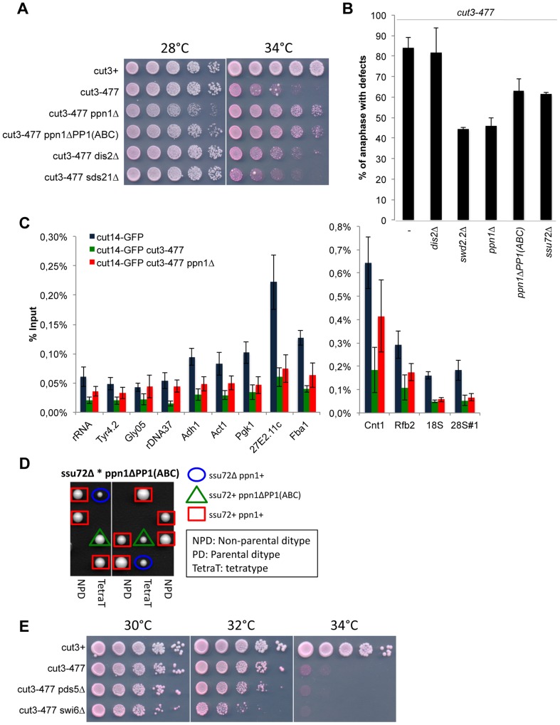
Functional links connecting gene transcription and condensin-mediated chromosome condensation have been established in species ranging from prokaryotes to vertebrates. However, the exact nature of these links remains misunderstood. Here we show in fission yeast that the 3' end RNA processing factor Swd2.2, a component of the Cleavage and Polyadenylation Factor (CPF), is a negative regulator of condensin-mediated chromosome condensation. Lack of Swd2.2 does not affect the assembly of the CPF but reduces its association with chromatin. This causes only limited, context-dependent effects on gene expression and transcription termination. However, CPF-associated Swd2.2 is required for the association of Protein Phosphatase 1 PP1(Dis2) with chromatin, through an interaction with Ppn1, a protein that we identify as the fission yeast homologue of vertebrate PNUTS. We demonstrate that Swd2.2, Ppn1 and PP1Dis2 form an independent module within the CPF, which provides an essential function in the absence of the CPF-associated Ssu72 phosphatase. We show that Ppn1 and Ssu72, like Swd2.2, are also negative regulators of condensin-mediated chromosome condensation. We conclude that Swd2.2 opposes condensin-mediated chromosome condensation by facilitating the function of the two CPF-associated phosphatases PP1 and Ssu72.


Conflict of interest statement

The authors have declared that no competing interests exist.

Figures

Figure 1
Figure 1. Swd2.2 antagonizes the association of Condensin with chromatin.
A. Serial dilutions of the indicated strains were plated on rich media at the indicated temperatures. B. Chromosome segregation in anaphase was visualized after tubulin staining in cells of the indicated genotypes grown for one generation at 34°C. C. Anaphases were scored as defective when chromatin was detected lagging between the two main DNA masses. For each genotype, a minimum of 6 independent experiments was performed in which a minimum of 100 anaphase cells was scored. ***p<0,001 Wilcoxon - Mann Whitney. D and E. Same as A and C, except that in E, 3 independent experiments were performed. F. The indicated strains were grown at 34°C for 3 hours and ChIP-qPCR was performed to analyze the amount of Cut14-GFP cross-linked to chromatin (mean ± standard deviation from 6 biological replicates). See text for details for the statistical analysis of the experiments.
Figure 2
Figure 2. Lack of Swd2.2 reduces the association of the RNA Pol III transcription machinery with chromatin.
A. Asynchronous populations of the indicated strains were grown at 30°C and ChIP-qPCR was performed to analyze the amount of Swd2.2-3flag cross-linked to chromatin (mean ± standard deviation from 4 biological replicates). BC. ChIP qPCR analysis of the indicated strains grown at 30°C. Mean ± standard deviation from 5 biological replicates. *<0,05; **<0,01; Wilcoxon - Mann Whitney. D. Western blot analysis of total protein extracts of the indicated strains. Tubulin (TAT1 antibody) is used as a loading control. E. qRT-PCR analysis of sfc6 expression in swd2.2+ and swd2.2Δ cells (mean ± standard deviation from 3 biological replicates) F. Serial dilutions of the indicated strains were plated on rich media at the indicated temperatures.
Figure 3
Figure 3. Swd2.2 facilitates the function of the CPF.
A. Serial dilutions of the indicated strains were plated on rich media at the indicated temperatures. B. The CPF component Yth1 tagged at the endogenous locus with 3flag epitopes was immuno-precipitated from cycling cells in the presence or absence of Swd2.2. Whole cell extracts (WCE) and the immuno-precipitated material (Flag IP) were analyzed by western blot. Yth1-3flag interacts with Pfs2-GFP whether or not Swd2.2 is present. Arrows indicate aspecific bands on the western blot. C. Asynchronous populations of the indicated strains were grown at 30°C and ChIP-qPCR was performed to analyze the amount of Pfs2-GFP cross-linked to chromatin (mean ± standard deviation from 4 biological replicates). D. Western blot analysis of total protein extracts of the indicated strains. Tubulin (TAT1 antibody) is used as a loading control.
Figure 4
Figure 4. Swd2.2 facilitates the localization of PP1 phosphatase by interacting with the PNUTS homologue Ppn1.
A. The interaction of Ppn1 with PP1Dis2 requires the three PP1-binding sites of Ppn1. GFP-tagged PP1Dis2 was immuno-precipitated from cycling cells in the presence of Flag-tagged Ppn1 (Ppn1-3flag) or Flag-tagged Ppn1 lacking the three PP1-binding sites (ppn1ΔPP1(ABC)-3flag). Whole cell extracts (WCE) and the immuno-precipitated material (GFP IP) were analyzed by western blot. B. Asynchronous populations of the indicated strains (Ppn1-3flag or GFP-PP1Dis2) were grown at 30°C and ChIP-qPCR was performed to analyze their enrichment at various sites along chromosomes. Enrichments were normalized to the values obtained at the RNA Polymerase I-transcribed 18S (mean ± standard deviation from 3 biological replicates). C. Lack of Ppn1 disrupts the nuclear localisation of GFP-tagged PP1Dis2. GFP-PP1Dis2 was imaged in dividing cells co-expressing (left panel) the mCherry-tagged nuclear envelope marker Mlp1 or (right panel) the mRFP-tagged nucleolar marker Fib1. DE. Asynchronous populations of the indicated strains were grown at 30°C and ChIP-qPCR was performed (mean ± standard deviation from 6 biological replicates). F. The protein stability of GFP-PP1Dis2 was assessed by western blot in the various mutant backgrounds used in DE. Tubulin was used as a loading control. G. The interaction between Flag-tagged Swd2.2 and Pk-tagged PP1Dis2 was analyzed by co-immunoprecipitation in protein extracts prepared from cycling cells in the presence or absence of Ppn1. Protein extracts were treated or not with RNase A prior to immuno-precipitation. Whole cell extracts (WCE) and the immuno-precipitated material (GFP IP) were analyzed by western blot. H. Asynchronous populations of the indicated strains were grown at 30°C and ChIP-qPCR was performed to analyze their enrichment at various sites along chromosomes. I. The protein stability of Flag-tagged Swd2.2 in the presence or absence of Ppn1 was assessed by western blot. Tubulin was used as a loading control. J. Asynchronous populations of the indicated strains were grown at 30°C. Protein extracts were prepared and analyzed by western blot using the indicated antibodies.
Figure 5
Figure 5. Swd2.2, Ppn1 and PP1Dis2 associate as a protein module to the CPF.
A. The interaction between Flag-tagged Swd2.2 and GFP-tagged Pfs2 was analyzed by co-immunoprecipitation in protein extracts prepared from cycling cells in the presence or absence of Ppn1. Whole cell extracts (WCE) and the immuno-precipitated material (GFP IP) were analyzed by western blot. BCDE. The proteins indicated at the top were purified by affinity and their associated partners were identified by MS/MS mass-spectrometry analysis (see Methods). The number of unique peptides recovered for each protein is indicated. F. Scheme summarizing the proteomic data. GH. Cells expressing the indicated epitope-tagged proteins were synchronized in early mitosis (mitotic) or not (cycling), using the cold-sensitive nda3KM311 mutation . G. Flag-tagged Swd2.2 was immuno-precipitated to look at its interaction with the core CPF component Pfs2. H. Flag-tagged PP1Dis2 was immuno-precipitated to look at its interaction with the DPS component Swd2.2.
Figure 6
Figure 6. Ppn1 and Ssu72 oppose condensin-mediated chromosome condensation.
A. Serial dilutions of the indicated strains were plated on rich media at the indicated temperatures. B. Chromosome segregation in anaphase was visualized after growing cells of the indicated genotypes for one generation at 34°C. Anaphases were scored as defective when lagging chromatin was detected between the two main DNA masses. For each genotype, a minimum of 6 independent experiments was performed in which a minimum of 100 anaphase cells was scored. C. The indicated strains were grown at 34°C for 3 hours and ChIP-qPCR was performed to analyze the amount of Cut14-GFP cross-linked to chromatin (mean ± standard deviation from 8 biological replicates). See text for details for the statistical analysis of the experiments. D. Tetrad dissection was used to show that the double mutant ssu72Δ ppn1ΔPP1(ABC) is dead. E. Serial dilutions of the indicated strains were plated on rich media at the indicated temperatures.

Similar articles

Cited by

References

    1. Piazza I, Haering CH, Rutkowska A (2013) Condensin: crafting the chromosome landscape. Chromosoma 122: 175–190. - PubMed
    1. Tada K, Susumu H, Sakuno T, Watanabe Y (2011) Condensin association with histone H2A shapes mitotic chromosomes. Nature 474: 477–483. - PubMed
    1. D'Ambrosio C, Schmidt CK, Katou Y, Kelly G, Itoh T, et al. (2008) Identification of cis-acting sites for condensin loading onto budding yeast chromosomes. Genes Dev 22: 2215–2227. - PMC - PubMed
    1. Kim JH, Zhang T, Wong NC, Davidson N, Maksimovic J, et al. (2013) Condensin I associates with structural and gene regulatory regions in vertebrate chromosomes. Nat Commun 4: 2537. - PMC - PubMed
    1. Tanaka A, Tanizawa H, Sriswasdi S, Iwasaki O, Chatterjee AG, et al. (2012) Epigenetic regulation of condensin-mediated genome organization during the cell cycle and upon DNA damage through histone H3 lysine 56 acetylation. Mol Cell 48: 532–546. - PMC - PubMed

Publication types

MeSH terms