. 2014 Jun 19;9(6):e100392.
doi: 10.1371/journal.pone.0100392. eCollection 2014.

Vaccine safety and efficacy evaluation of a recombinant bovine respiratory syncytial virus (BRSV) with deletion of the SH gene and subunit vaccines based on recombinant human RSV proteins: N-nanorings, P and M2-1, in calves with maternal antibodies

Affiliations

Vaccine safety and efficacy evaluation of a recombinant bovine respiratory syncytial virus (BRSV) with deletion of the SH gene and subunit vaccines based on recombinant human RSV proteins: N-nanorings, P and M2-1, in calves with maternal antibodies

Krister Blodörn et al. PLoS One. .

Abstract

The development of safe and effective vaccines against both bovine and human respiratory syncytial viruses (BRSV, HRSV) to be used in the presence of RSV-specific maternally-derived antibodies (MDA) remains a high priority in human and veterinary medicine. Herein, we present safety and efficacy results from a virulent BRSV challenge of calves with MDA, which were immunized with one of three vaccine candidates that allow serological differentiation of infected from vaccinated animals (DIVA): an SH gene-deleted recombinant BRSV (ΔSHrBRSV), and two subunit (SU) formulations based on HRSV-P, -M2-1, and -N recombinant proteins displaying BRSV-F and -G epitopes, adjuvanted by either oil emulsion (Montanide ISA71VG, SUMont) or immunostimulating complex matrices (AbISCO-300, SUAbis). Whereas all control animals developed severe respiratory disease and shed high levels of virus following BRSV challenge, ΔSHrBRSV-immunized calves demonstrated almost complete clinical and virological protection five weeks after a single intranasal vaccination. Although mucosal vaccination with ΔSHrBRSV failed to induce a detectable immunological response, there was a rapid and strong anamnestic mucosal BRSV-specific IgA, virus neutralizing antibody and local T cell response following challenge with virulent BRSV. Calves immunized twice intramuscularly, three weeks apart with SUMont were also well protected two weeks after boost. The protection was not as pronounced as that in ΔSHrBRSV-immunized animals, but superior to those immunized twice subcutaneously three weeks apart with SUAbis. Antibody responses induced by the subunit vaccines were non-neutralizing and not directed against BRSV F or G proteins. When formulated as SUMont but not as SUAbis, the HRSV N, P and M2-1 proteins induced strong systemic cross-protective cell-mediated immune responses detectable already after priming. ΔSHrBRSV and SUMont are two promising DIVA-compatible vaccines, apparently inducing protection by different immune responses that were influenced by vaccine-composition, immunization route and regimen.


Conflict of interest statement

Competing Interests: The authors have read the journal’s policy and have the following conflicts: Jean-François Eléouët and Sabine Riffault (co-inventors) hold two patents on N-rings usage for RSV vaccination (WO/2006/117456) and N-rings as immunogenic carrier for heterologous antigens (WO/2007/119011). These patents belong to their institution INRA and this does not alter the authors’ adherence to all PLOS ONE policies on sharing data and materials.

Figures

Figure 1
Figure 1. Experiment timeline, vaccination and sampling.
Twenty calves with moderate titers of BRSV-specific serum antibodies (MDA) were allocated into 4 groups and vaccinated as indicated in the figure; all were vaccinated on post-vaccination day (PVD) 0 (Vacc. I, white arrow) with either (a) 5×10⁶ pfu of ΔSHrBRSV intranasally (i.n.); (b) BRSV and HRSV recombinant protein subunits (SU) adjuvanted by Montanide (SUMont) intramuscularly (i.m.), (c) SU adjuvanted by AbISCO-300 (SUAbis) subcutaneously (s.c.), or (d) adjuvant alone s.c. (Controls). On PVD 20, all animals except those immunized with ΔSHrBRSV, were boosted with the same formulation and route as for Vacc. I (Vacc. II, gray arrow). Three BRSV-seronegative calves were housed in contact with ΔSHrBRSV-infected animals to determine transmission of the vaccine virus (Sentinel calves), and monitored until euthanized (†) on PVD 22. On PVD 20, one calf in group c was euthanized due to traumatic injury. On post-infection day (PID) 0, all calves were challenged i.n. with 104 pfu virulent BRSV (black arrow), and clinically scored daily until PID 7. Throughout the experiment, samples were collected, as indicated in the figure, to analyze antibodies in serum and nasal secretions, ex-vivo response of peripheral blood mononuclear cells (PBMC) to restimulation with BRSV, and virus shedding in nasal secretions (Nasal swab). At post-mortem examination (PM), lung lesions were recorded and tissue samples collected, as well as bronchoalveolar lavage (BAL) samples for antibody, BRSV RT-PCR and virus isolation.
Figure 2
Figure 2. Vaccination protects against clinical signs of BRSV disease.
Four groups of 5 calves were vaccinated as described in Fig. 1 and challenged with BRSV, 5 weeks after vaccination, on post-infection day (PID) 0. Following challenge, calves were examined daily until euthanization on PID 7, and the severity of clinical signs of diseases were scored as previously described . (A) presents the mean square root of clinical scores per day (to approximate normal distribution for statistical analysis), and (B) the accumulated clinical score from PID 0 to PID 7, with standard deviations indicated by upward deflecting lines. Statistically significant differences are indicated by asterisks p≤0.05 (*); p≤0.01 (**); p≤0.005 (***); p≤0.001 (****); p≤0.0001 (*****).
Figure 3
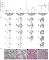
Figure 3. Vaccination reduces the extent of lung lesions following BRSV challenge.
Four groups of 5 calves were vaccinated as described in Fig. 1 and challenged with BRSV, 5 weeks after vaccination. Two weeks before challenge, one calf (c5) was euthanized due to traumatic injury. Lungs were removed after exsanguination, lesions were recorded on a lung chart after visual examination and palpation, and the proportion of lung showing pneumonic consolidation was calculated. Formalin-fixed tissue samples from each lobe in the right lung were analyzed for the severity of histopathological changes and scored as either normal (0), mild (1), moderate (2) or severe (3). (A) shows the extent of macroscopic lesions on the y-axes, and the microscopic severity of inflammation (mean score of four sections per calf) on the x-axes. Statistically significant difference is indicated by asterisks (p≤0.05). (B) shows the percent of pneumonic consolidation in each animal (also depicted as filled areas in lung-charts), and emphysema (outlined areas in calves d3 and d4). Panels C (I–IV) show representative histological images from each of the four groups of calves. Bar indicate 100 µm. Panels C (I) (ΔSHrBRSV), C (II) (SUMont), C (III) (SUAbis) and C (IV) (Control) show lung parenchyma with minimal, mild, moderate and severe pathological changes, respectively.
Figure 4
Figure 4. Vaccination reduces virus load in upper and lower airways following virulent BRSV challenge.
Four groups of 5 calves were vaccinated as described in Fig. 1 and challenged with BRSV, 5 weeks after vaccination, on post-infection day (PID) 0. Two weeks before challenge, one calf (c5) was euthanized due to traumatic injury. The figure presents mean viral load in nasal swabs collected from PID 0 to PID 7 in panel A and post-mortem bronchoalveolar lavage (BAL) in panel B, as determined by BRSV F-gene RT-PCR after total RNA extraction, and is expressed as TCID50 equivalent, calculated from standard dilution series of virus with a known TCID50. The area under mean curves in panel A represents the accumulated detected virus shed (AVS): calves immunized with either ΔSHrBRSV or SUMont had significantly lower AVS (1.4±2.2 eqTCID50, p≤0.005 and 3.6±2.6 eqTCID50, p≤0.05 respectively), compared to calves immunized with either SUAbis (7.9±3.4 eqTCID50) or adjuvant alone (11.0±2.2 eqTCID50). Statistically significant difference with Student’s t-test are indicated by asterisks and the corresponding groups; p≤0.05 (*); p≤0.01 (**); p≤0.005 (***); p≤0.001 (****).
Figure 5
Figure 5. Vaccination reduces clinical signs, lung pathology and viral replication, following virulent BRSV challenge.
Four groups of 5 calves were vaccinated as described in Fig. 1 and challenged with BRSV, 5 weeks after vaccination. Two weeks before challenge, one calf (c5) was euthanized due to traumatic injury. Calves were ranked (1–19) in each of three post-challenge parameters, with a high clinical rank indicating a high accumulated clinical score (Fig. 2); a high lung lesion rank indicating a high percent of macroscopic lung lesions post-mortem (Fig. 3); and a high viral-shed rank indicating a high accumulated viral-shed following challenge (Fig. 4). The figure shows the group sum of each of these ranks. To correct for the unequal number of calves per group (n), each rank sum was divided by n, and multiplied by 5. The stacked bars per group represent the sum of rank sums (total rank sum). Statistically significant differences in individual rank sums are indicated by asterisks and the corresponding group; p≤0.05 (*); p≤0.001 (****).
Figure 6
Figure 6. RSV-specific serum antibodies in calves before and after immunization and subsequent challenge with virulent BRSV.
Four groups of 5 calves were vaccinated as described in Fig. 1 (white and grey arrows) and challenged with BRSV, 5 weeks after vaccination (black arrow) on post-infection day (PID) 0. Two weeks before challenge, one calf (C5) was euthanized due to traumatic injury. Panels show group mean log10 serum titers of: (A) BRSV-specific IgG1 (by ELISA); (B) IgG directed against BRSV G on PID 0 and PID 7 (by ELISA); (C) IgG directed against HRSV F on PID 0 and PID 7 (by ELISA); (D) IgG directed against HRSV N (by ELISA); (E) IgG directed against HRSV P (by ELISA); and (F) IgG directed against HRSV M2-1 (by ELISA) (G) BRSV-neutralizing antibodies (by plaque reduction assay). Note that the scale of the y-axis is not uniform between panels. Statistically significant difference on PID 7 is indicated by asterisks and the corresponding group; p≤0.05 (*); p≤0.01 (**); p≤0.005 (***); p≤0.001 (****).
Figure 7
Figure 7. Mucosal IgA antibodies in the upper and lower airways, before and after BRSV challenge.
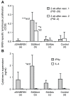
Four groups of 5 calves were vaccinated as described in Fig. 1 and challenged with BRSV, 5 weeks after vaccination. Two weeks before challenge, one calf (c5) was euthanized due to traumatic injury. BRSV-specific IgA antibodies were analyzed by ELISA. (A) shows group mean levels of BRSV-specific IgA in nasal secretions on post-infection day (PID) 0 and 7, whereas (B) and (C) show group mean titers of total BRSV- and HRSV-N-specific IgA in bronchoalveolar lavage (BAL) on PID 7, respectively. BAL samples were titrated, whereas antibody levels in nasal secretions were semi-quantitatively determined and expressed as a percentage of a positive control sample, due to lack of sample material. Standard deviations are indicated by upward deflecting lines. Statistically significant differences between PID 0 and PID 7 in panel A are indicated by a horizontal line, whereas in all panels significant differences between groups for the same time-point are indicated by asterisks and the corresponding group letter; p≤0.05 (*); p≤0.01 (**); p≤0.005 (***); p≤0.001 (****).
Figure 8
Figure 8. BRSV-specific lymphocyte proliferative response in vaccinated calves.
Four groups of 5 calves were vaccinated as described in Fig. 1 and challenged with BRSV, 5 weeks after vaccination, on post-infection day (PID) 0. Two weeks before challenge, one calf (c5) was euthanized due to traumatic injury. Peripheral blood mononuclear cells (PBMC) were purified from blood two weeks after first and second vaccination, as indicated in Fig. 1, and stimulated ex-vivo with either BRSV-infected or uninfected cell lysate. (A) Corrected optical density (COD) of Alamar Blue (Invitrogen, Sweden), indicating proliferative response after seven days of incubation. (B) IFNγ and IL-4 in supernatant from PBMC restimulated with BRSV-infected cell lysate, expressed as group means (ng/ml). Standard deviations are indicated by upward deflecting lines. Statistically significant differences are indicated by asterisks and the corresponding group; p≤0.05 (*); p≤0.01 (**); p≤0.001 (****).

Similar articles

Cited by

References

    1. Valarcher J-F, Taylor G (2007) Bovine respiratory syncytial virus infection. Vet Res 38: 153–180 10.1051/vetres:2006053 - DOI - PubMed
    1. Seiffert SN, Hilty M, Perreten V, Endimiani A (2013) Extended-spectrum cephalosporin-resistant gram-negative organisms in livestock: An emerging problem for human health? Drug Resist Updat. doi:10.1016/j.drup.2012.12.001. - PubMed
    1. Fulton RW (2009) Bovine respiratory disease research (1983–2009). Anim Health Res Rev 10: 131–139 10.1017/S146625230999017X - DOI - PubMed
    1. Kimman TG, Zimmer GM, Westenbrink F, Mars J, van Leeuwen E (1988) Epidemiological study of bovine respiratory syncytial virus infections in calves: influence of maternal antibodies on the outcome of disease. Vet Rec 123: 104–109. - PubMed
    1. Kimman TG, Westenbrink F, Straver PJ (1989) Priming for local and systemic antibody memory responses to bovine respiratory syncytial virus: effect of amount of virus, virus replication, route of administration and maternal antibodies. Vet Immunol Immunopathol 22: 145–160. - PubMed

Publication types

MeSH terms