. 2014 Aug;95(4):271-81.
doi: 10.1111/iep.12085. Epub 2014 Jun 19.

Destruction of the hepatocyte junction by intercellular invasion of Leptospira causes jaundice in a hamster model of Weil's disease

Affiliations

Destruction of the hepatocyte junction by intercellular invasion of Leptospira causes jaundice in a hamster model of Weil's disease

Satoshi Miyahara et al. Int J Exp Pathol. 2014 Aug.

Abstract

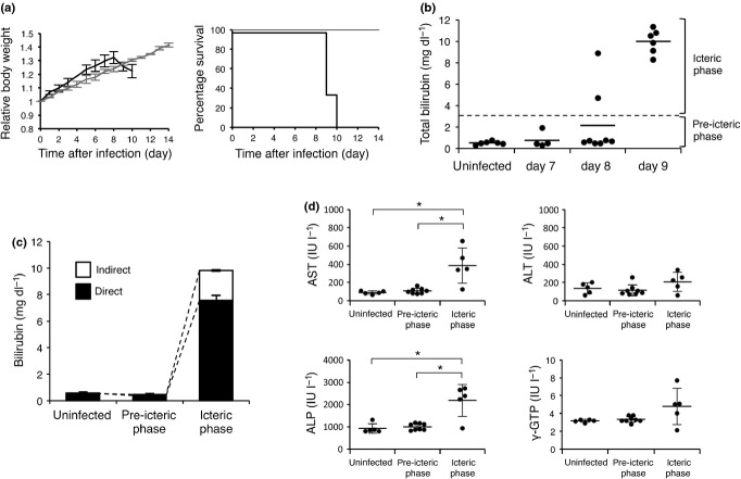
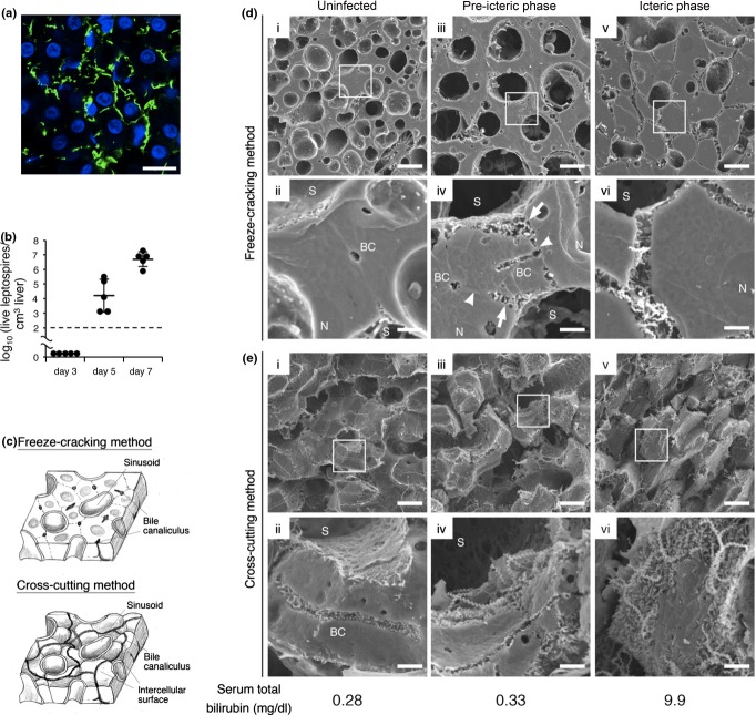
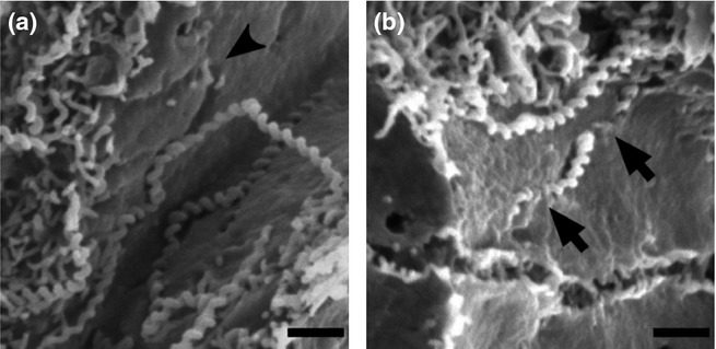
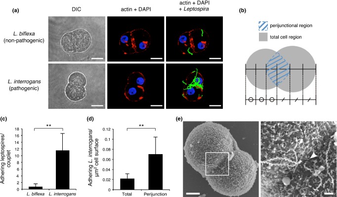
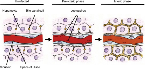
Weil's disease, the most severe form of leptospirosis, is characterized by jaundice, haemorrhage and renal failure. The mechanisms of jaundice caused by pathogenic Leptospira remain unclear. We therefore aimed to elucidate the mechanisms by integrating histopathological changes with serum biochemical abnormalities during the development of jaundice in a hamster model of Weil's disease. In this work, we obtained three-dimensional images of infected hamster livers using scanning electron microscope together with freeze-cracking and cross-cutting methods for sample preparation. The images displayed the corkscrew-shaped bacteria, which infiltrated the Disse's space, migrated between hepatocytes, detached the intercellular junctions and disrupted the bile canaliculi. Destruction of bile canaliculi coincided with the elevation of conjugated bilirubin, aspartate transaminase and alkaline phosphatase levels in serum, whereas serum alanine transaminase and γ-glutamyl transpeptidase levels increased slightly, but not significantly. We also found in ex vivo experiments that pathogenic, but not non-pathogenic leptospires, tend to adhere to the perijunctional region of hepatocyte couplets isolated from hamsters and initiate invasion of the intercellular junction within 1 h after co-incubation. Our results suggest that pathogenic leptospires invade the intercellular junctions of host hepatocytes, and this invasion contributes in the disruption of the junction. Subsequently, bile leaks from bile canaliculi and jaundice occurs immediately. Our findings revealed not only a novel pathogenicity of leptospires, but also a novel mechanism of jaundice induced by bacterial infection.

Keywords: Leptospira; bacterial invasion; cell junction; hepatocyte couplet; jaundice; scanning electron microscope.


Figures

Figure 1
Figure 1
Leptospira interrogans strain K37 infection causes sudden onset of jaundice and cholestasis-type abnormality of serum chemistries. (a) Relative body weight and survival of uninfected hamsters (n = 3, grey) or hamsters subcutaneously infected with L. interrogans strain K37 (n = 6, black). (b) Serum total bilirubin concentrations of uninfected (n = 6) or infected hamsters at seventh (n = 4), eighth (n = 8) or ninth (n = 6) day postinoculation. Horizontal dashed line indicates 3 mg/dl to distinguish pre-icteric and icteric phase. Horizontal bars indicate the mean. (c,d) Serum measurements of uninfected hamsters (n = 6) and infected hamsters, which were divided into two groups (pre-icteric phase; n = 10 and icteric phase; n = 8) according to serum bilirubin concentration. (c) Serum concentrations of direct and indirect bilirubin. (d) Serum concentrations of aspartate transaminase (AST), alanine transaminase (ALT), alkaline phosphatase (ALP) and γ-glutamyl transpeptidase (γ-GTP). Data are shown as the mean ± SD. Statistical significance is shown by *P < 0.05 as determined by Welch's t-test.
Figure 2
Figure 2
Disruption of intercellular adhesion caused by leptospires in the liver triggers jaundice. (a) Representative confocal images of liver from hamsters infected with Leptospira interrogans strain K37 at eighth day postinoculation showing leptospires stained with rabbit polyclonal antisera and Alexa Fluor 488 conjugated anti-rabbit monoclonal antibody (green), and nuclei of liver cells stained with DAPI (blue) Scale bar, 10 μm. (b) Numbers of culturable leptospires in 1 cm3 of hamster livers at third, fifth and seventh day postinoculation determined by dilution method. Black dashed line represents the limit of detection. Data are the mean ± SD. (c) Schematic diagram of the liver structure exposed by two methods of sample preparation. Freeze-cracking method exposes flat fractured surface (top image). Cross-cutting method exposes intercellular surface (bottom image). (d,e) Representative scanning electron microscope images of the livers from uninfected hamsters (i and ii) or infected hamsters in pre-icteric (iii and iv) or icteric phase (v and vi) using freeze-cracking (d) and cross-cutting methods (e). The framed areas in the top images are enlarged in the bottom images. The scale bars represent 10 μm for the top images and 2 μm for the bottom images. The arrows show hepatocyte membrane detached from the adjacent cell. The arrowheads point to hepatocyte membranes that remained attached to the adjoining cell. Total bilirubin concentration of each hamster is indicated below the images. BC, bile canaliculi; N, nucleus of the hepatocyte; and S, sinusoid.
Figure 3
Figure 3
Leptospira invade liver parenchyma by paracellular route. (a,b) Representative scanning electron microscope images of liver from infected hamsters showing intercellular surface of hepatocytes exposed by cross-cutting method. The black arrowhead shows the trail formed by a leptospire on the intercellular surface (a). The black arrows point to penetration by a leptospire through cell membrane (b). Scale bars, 1 μm.
Figure 4
Figure 4
Attachment and invasion to host hepatocytes are characteristic of pathogenic leptospires. (a) Representative confocal images of isolated hamster hepatocyte couplets infected with pathogenic (Leptospira interrogans) or non-pathogenic (Leptospira biflexa) leptospires on coverslips for 1 h showing actin accumulation stained with Acti-stain 555 phalloidin (red), and leptospires stained with rabbit polyclonal antisera and Alexa Fluor 488 conjugated anti-rabbit monoclonal antibody (green). Nuclei of liver cells were stained with DAPI (blue). Non-adherent bacteria were washed out. Projections of three-dimensional reconstructions are shown Scale bar, 10 μm. (b) Schematic diagram of hepatocyte couplet indicating the total cell (grey) and perijunctional (blue striped) regions. The perijunctional region was defined as an ellipse with its long axis overlying the junction of the two hepatocytes, and its transverse axis extending over one-third of the transverse diameter of each cell. The total cell region included the perijunctional region. (c,d) Analysis of the number of pathogenic or non-pathogenic leptospires adhering to the total cell region of a couplet (c) and the number of adhering pathogenic leptospires per μm2 of total cell or perijunctional regions (d). n = 50 per group. Data are shown as the mean ± SD. Statistical significance is shown by **P < 0.001 as determined by Welch's t-test. (e) Representative scanning electron microscope images of isolated hamster hepatocyte couplet infected by L. interrogans strain K37. The framed area in the left image is enlarged in the right image. The scale bar represents 5 μm for the left image and 1 μm for the right image. The white arrowhead shows one end of a spirochete entering the intercellular junction (right).
Figure 5
Figure 5
Schematic representation of jaundice caused by pathogenic Leptospira. In the normal state, a tight junction seals bile canaliculi and prevents leakage of bile to bloodstream (left). Once pathogenic leptospires reach the liver sinusoids, they have the property to attach to and invade intercellular junctions of hepatocytes and this invasion contributes to disrupt the junction (middle). When the tight junction is disrupted, bile leaks from bile canaliculi and this result in jaundice (right).

References

    1. Anderson JM. Leaky junctions and cholestasis: a tight correlation. Gastroenterology. 1996;110:1662–1665. - PubMed
    1. Arean VM. The pathologic anatomy and pathogenesis of fatal human leptospirosis (Weil's disease) Am. J. Pathol. 1962a;40:393–423. - PMC - PubMed
    1. Arean VM. Studies on the pathogenesis of leptospirosis. II. A clinicopathologic evaluation of hepatic and renal function in experimental leptospiral infections. Lab. Invest. 1962b;11:273–288. - PubMed
    1. Balkovetz DF, Katz J. Bacterial invasion by a paracellular route: divide and conquer. Microbes Infect. 2003;5:613–619. - PubMed
    1. Ballard SA, Williamson M, Adler B, Vinh T, Faine S. Interaction of virulent and avirulent leptospires with primary cultures of renal epithelial cells. J. Med. Microbiol. 1986;21:59–67. - PubMed

Publication types

MeSH terms

LinkOut - more resources