. 2014 Jun 19;10(6):e1004197.
doi: 10.1371/journal.ppat.1004197. eCollection 2014 Jun.

The calcium-dependent protein kinase 3 of toxoplasma influences basal calcium levels and functions beyond egress as revealed by quantitative phosphoproteome analysis

Affiliations

The calcium-dependent protein kinase 3 of toxoplasma influences basal calcium levels and functions beyond egress as revealed by quantitative phosphoproteome analysis

Moritz Treeck et al. PLoS Pathog. .

Abstract

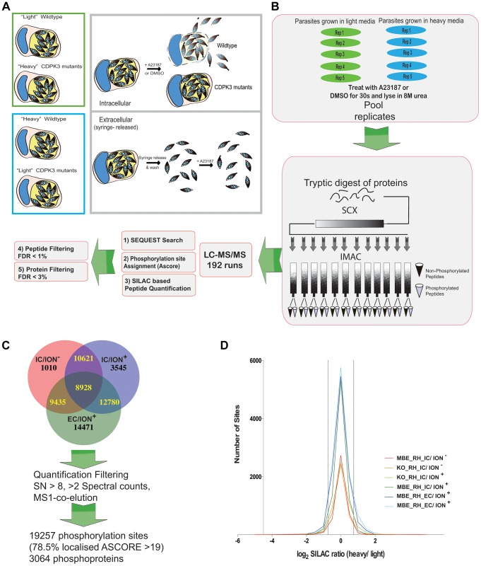
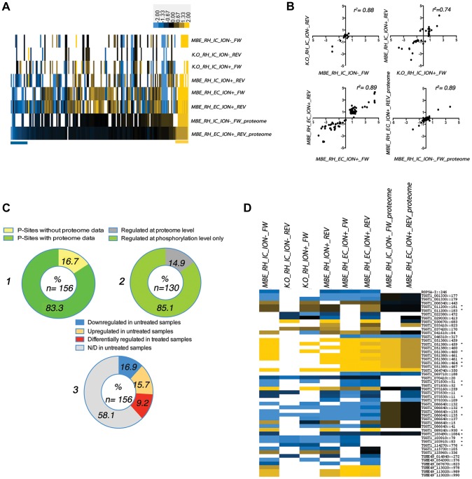
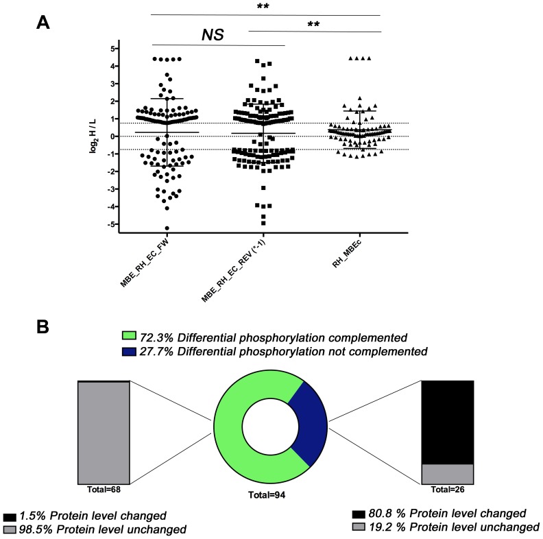
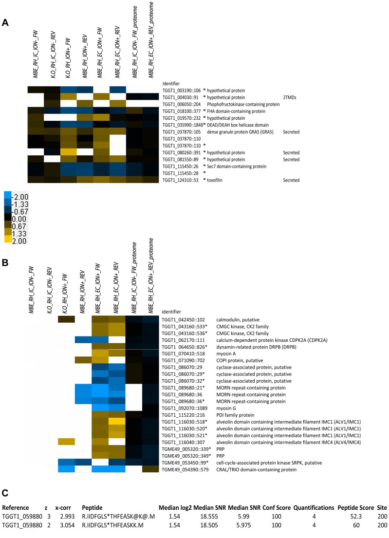
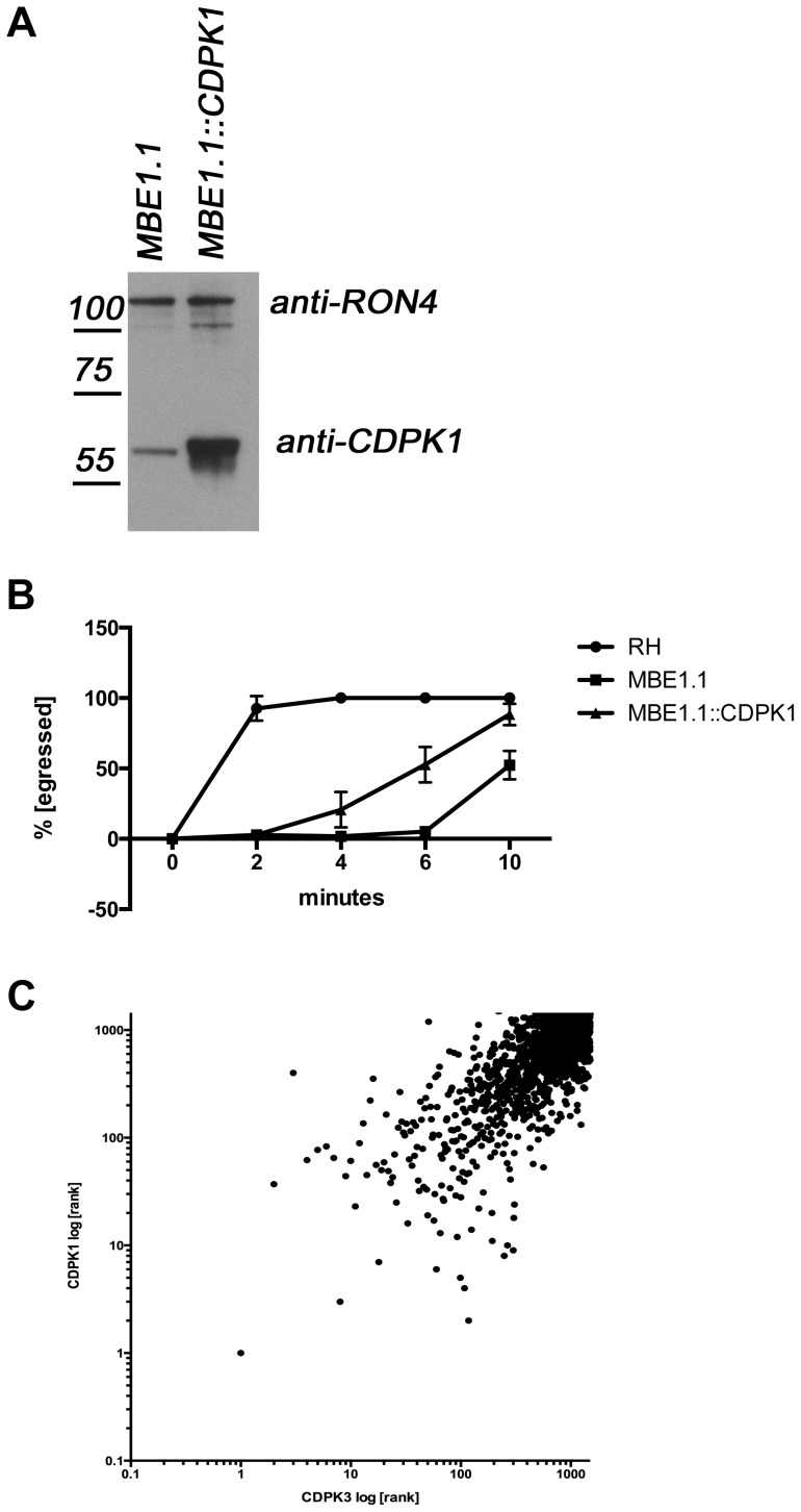
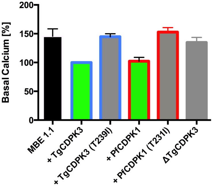
Calcium-dependent protein kinases (CDPKs) are conserved in plants and apicomplexan parasites. In Toxoplasma gondii, TgCDPK3 regulates parasite egress from the host cell in the presence of a calcium-ionophore. The targets and the pathways that the kinase controls, however, are not known. To identify pathways regulated by TgCDPK3, we measured relative phosphorylation site usage in wild type and TgCDPK3 mutant and knock-out parasites by quantitative mass-spectrometry using stable isotope-labeling with amino acids in cell culture (SILAC). This revealed known and novel phosphorylation events on proteins predicted to play a role in host-cell egress, but also a novel function of TgCDPK3 as an upstream regulator of other calcium-dependent signaling pathways, as we also identified proteins that are differentially phosphorylated prior to egress, including proteins important for ion-homeostasis and metabolism. This observation is supported by the observation that basal calcium levels are increased in parasites where TgCDPK3 has been inactivated. Most of the differential phosphorylation observed in CDPK3 mutants is rescued by complementation of the mutants with a wild type copy of TgCDPK3. Lastly, the TgCDPK3 mutants showed hyperphosphorylation of two targets of a related calcium-dependent kinase (TgCDPK1), as well as TgCDPK1 itself, indicating that this latter kinase appears to play a role downstream of TgCDPK3 function. Overexpression of TgCDPK1 partially rescues the egress phenotype of the TgCDPK3 mutants, reinforcing this conclusion. These results show that TgCDPK3 plays a pivotal role in regulating tachyzoite functions including, but not limited to, egress.


Conflict of interest statement

The authors have declared that no competing interests exist.

Figures

Figure 1
Figure 1. Generation of phosphoproteome samples.
A) Illustration of the experimental conditions used in this study and the functional consequences of treatment with the calcium ionophore A23187. Upper panels show “intracellular” conditions: infected cells containing mutant or wild type parasites were labeled with “heavy” or “light” SILAC media, treated with A23187 ionophore or DMSO for 30 sec and subsequently lysed for phosphoproteome analysis. Whereas wildtype parasites rapidly egress upon ionophore treatment (after 2 minutes, 100% have exited the host cell), TgCDPK3 mutant parasites extrude the conoid, a sign that they have sensed the ionophore, but fail to egress in a timely matter. Lower panels show “extracellular” conditions: all as for “intracellular” except the cultures were scraped and passed through a syringe to release the parasites followed by ionophore treatment for 30 seconds. All conditions were performed in “forward” and “reverse” (where the heavy and light labeling are swapped). B) 5 dishes (“rep”) per condition (or 5 vials containing extracellular parasites) were individually treated for 30 seconds prior to lysis, mixing, reduction and alkylation. SCX chromatography and IMAC were used for phosphopeptide enrichment. Blue and green color-coding represents parasites grown in “light” or “heavy” media, respectively. In total, 96 samples were analyzed in duplicate by LC-MS/MS followed by bioinformatic filtering to reduce the site and protein FDR to <1% and 3%, respectively. C) A total of 32,147 phosphorylation sites were identified in the three datasets: intracellular treated with ionophore (IC/ION+) or DMSO (IC/ION) and extracellular treated with ionophore (EC/ION+). Further filtering based on criteria specified in the text, including signal/noise ratio >8 (SN>8) reduced the set of quantified phosphorylation sites to 19,257 in 3064 proteins (See also Table S1). D) Histogram of median-centered log2 H/L SILAC ratios for each dataset comparing a TgCDPK3 mutant and wild type RH. MBE indicates the MBE1.1 mutant that is resistant to the ionophore and KO indicates the TgCDPK3 knock-out mutant. The conditions are as described above.
Figure 2
Figure 2. Analysis of differential phosphorylation site usage and protein abundance in WT and TgCDPK3 mutant parasites (see Figure 1 and Figure S1 for a description of experimental conditions).
A) 156 phosphorylation sites were identified as different between WT (RH) and TgCDPK3 mutant parasites under one or more of the conditions described in Fig. 1. FW = forward; REV = reverse labeling. The heat-map represents the log2 H/L SILAC ratios for the phosphoproteome or, where indicated, the proteome experiments. Each bar represents a phosphorylation site. Missing values are white. Only phosphorylation sites for which protein levels were also measured are displayed. A positive log2 score indicates higher abundance of the phosphopeptide in mutant compared to WT (RH) parasites. The results are sorted first for increasing SILAC ratios on the protein level (see also Table S2). Bars at the bottom of the heat map indicate phosphorylation sites where the difference in phosphorylation state can be explained by differences on the protein level. The color bar represents log2 fold-changes. B) Pearson correlation of phosphorylation sites and protein SILAC-ratios between WT (RH) and TgCDPK3 mutants identified in each condition (forward and reverse for “IC/ION”, “IC/ION+” and “EC/ION+”). Each dot represents one phosphosite and the log2 ratio under any one condition but with forward labeling being one mutant relative to WT and the reverse labeling being the other mutant relative to WT. C) 1) Percentage of the 156 differing phosphosites for which proteome information was also obtained; 2) Percentage of the 130 differing phosphosites for which proteome information was also obtained where the differences in the protein's abundance was (“Regulated at proteome level”) or was not (“Regulated at phosphorylation level only”) sufficient to explain the difference in the abundance of the phosphorylated peptide; 3) Distribution of phosphorylation sites regulated in the absence or presence of ionophore. Blue and yellow indicate the percentage of sites that appear less or more phosphorylated in the mutant samples relative to wild type in the “IC/ION“ conditions (“untreated”). Red indicates the percentage of sites that were identified as differentially phosphorylated in the “IC/ION+” conditions, but phosphorylated at similar levels between WT and mutant parasites in the “IC/ION“ conditions. Grey indicates phosphopeptides that were not detected (N/D) in the untreated samples. D) Heat-map showing all phosphorylation sites and proteins with changing phosphorylation ratios in “IC/ION”conditions, sorted by annotated gene identifier based on ToxoDB (v7.3). Samples are annotated as in Fig. 2A. The numbers next to the annotation indicate the position of the phosphorylation site within the protein. An asterisk (*) following the gene ID indicates quantifications are based on the bis-phosphorylated peptide form. The heat-map color intensities correspond to Figure 2A.
Figure 3
Figure 3. Phosphoproteome and proteome analysis of MBE1.1 complemented with wild type TgCDPK3.
A) All log2 H/L SILAC ratios for the 156 phosphorylation sites identified as differentially phosphorylated between TgCDPK3 mutants and wild type parasites are plotted from both extracellular phosphoproteome experiments and the corresponding SILAC ratios from the comparison of WT (RH) and the MBE1.1::CDPK3 complemented mutant (MBEc). Each point represents an individual phosphosite. The distribution of the SILAC ratios was not significantly different (NS) between the forward and the reverse experiments where MBE1.1 and RH parasites were compared. The RH vs. MBEc comparison showed most phosphosites were not significantly different between these two samples (log2 H/L values between 0.75 and −0.75) and the overall dataset was significantly different relative to the two comparing RH and the MBE1.1 mutant (**; p<0.001). B) Pie/bar charts showing that the majority of phosphorylation sites are complemented. Of the 156 TgCDPK3-dependent sites described in Fig. 2A, 94 gave reliable data in the comparison between the complemented mutant and WT strains. The central pie chart shows the percentage of these that were rescued by complementation (i.e., were no longer different relative to RH (WT); black) or were not rescued (i.e., still differed relative to WT; grey). The left bar shows that of the 68 (72.3%) sites that were complemented, only 1 (1.5%) was also changed in regards to the overall protein abundance. In contrast, a majority (20 or 80.1%) of the 26 phosphosites that were not rescued by complementation differed in protein abundance between WT and mutant (right bar).
Figure 4
Figure 4. Heat-map of phosphorylation sites regulated during egress.
A) Comparison of phosphorylation sites that show an ionophore-dependent difference between WT vs. mutant TgCDPK3 only when treated with ionophore, but equal phosphorylation site usage in the absence of ionophore. Conditions are as for Fig. 1 and 2. The color bars represent log2 H/L fold-changes for each. Identifier indicates the gene annotation and location of the site within the protein. An asterisk indicates whether a phosphorylation site has been identified on a bis-phosphorylated peptide. B) Heat-map of phosphorylation sites suspected or known to be involved in egress or motility. Details as for Part A except the phosphosites shown are all those that are not identified in the absence of ionophore and different when treated and were identified on proteins implicated in egress or motility. C) Phosphopeptide isoforms identified and quantified in this study containing the autophosphorylation (T200) from TgCDPK1. Median values for all peptide measurements are shown. The phosphorylated residue is marked (*).
Figure 5
Figure 5. Overexpression of TgCDPK1 partially rescues the egress defect.
A) TgCDPK1 expression in engineered parasites. Lysates from MBE1.1 parasites with and without TgCDPK1-HA expressed off the GRA2 promoter were separated by gel electrophoresis and blotted. Antibodies against TgCDPK1 were used to detect both the endogenous TgCDPK1 and TgCDPK1-HA expression (the endogenous protein migrates slightly faster than the HA-tagged version). The mass (in kDa) and migration of size markers are shown to the left RON4 expression was detected by blotting with rabbit anti-RON4 and served as a loading control. B) Overexpression of TgCDPK1 partially rescues egress. Parasites were treated with 1 µM A23187 and egress monitored over 10 minutes. Wild type (RH), TgCDPK3 mutant (MBE1.1) and MBE1.1 over-expressing TgCDPK1 (MBE1.1::CDPK1) were compared. C) TgCDPK1 and TgCDPK3 have overlapping substrate preferences. Peptide microarrays spotted with random 13-mer peptides with a central serine residue have been incubated with recombinant TgCDPK1 or TgCDPK1 and 32P-ATP. All peptides where ranked according to the ability of the respective kinase to phosphorylate any given peptide, with the highest phosphorylation ranked 1st. The scale is log10.
Figure 6
Figure 6. Absence of TgCDPK3 results in elevation of resting Ca2+ levels in T. gondii tachyzoites.
Ca2+ levels in Tgcdpk3 mutants (MBE 1.1), Tgcdpk3 mutants complemented with either wild-type or kinase mutant versions of TgCDPK3 (T239I), ΔTgCDPK3 and Tgcdpk3 mutants complemented with either wild-type or kinase mutant versions (T231I) of PfCDPK1 parasites was measured using the cell permeant calcium indicator Fluo-4-AM. Cell lines defective in egress are shown in grey, cell lines complemented with either WT TgCDPK3 or PfCDPK1 are capable of egress and are shown in green. Blue outlines show MBE1.1 complemented with TgCDPK3 variants while red outlines show data from MBE1.1 complemented with PfCDPK1 variants. Fluorescence values of MBE 1.1+ TgCDPK3 were set at 100% for relative comparison. n = 3 independent experiments. Error bars, SEM (** P<0.01, one-way ANOVA). ns =  not significant.

References

    1. Billker O, Lourido S, Sibley LD (2009) Calcium-dependent signaling and kinases in apicomplexan parasites. Cell Host Microbe 5: 612–622. - PMC - PubMed
    1. Billker O, Dechamps S, Tewari R, Wenig G, Franke-Fayard B, et al. (2004) Calcium and a calcium-dependent protein kinase regulate gamete formation and mosquito transmission in a malaria parasite. Cell 117: 503–514. - PubMed
    1. Kato N, Sakata T, Breton G, Le Roch KG, Nagle A, et al. (2008) Gene expression signatures and small-molecule compounds link a protein kinase to Plasmodium falciparum motility. Nat Chem Biol 4: 347–356. - PMC - PubMed
    1. Dvorin JD, Martyn DC, Patel SD, Grimley JS, Collins CR, et al. (2010) A plant-like kinase in Plasmodium falciparum regulates parasite egress from erythrocytes. Science 328: 910–912. - PMC - PubMed
    1. Lourido S, Shuman J, Zhang C, Shokat KM, Hui R, et al. (2010) Calcium-dependent protein kinase 1 is an essential regulator of exocytosis in Toxoplasma. Nature 465: 359–362. - PMC - PubMed

Publication types

LinkOut - more resources