. 2014 Jun 18;82(6):1255-1262.
doi: 10.1016/j.neuron.2014.04.036.

Off-target effect of doublecortin family shRNA on neuronal migration associated with endogenous microRNA dysregulation

Affiliations

Off-target effect of doublecortin family shRNA on neuronal migration associated with endogenous microRNA dysregulation

Seung Tae Baek et al. Neuron. .

Abstract

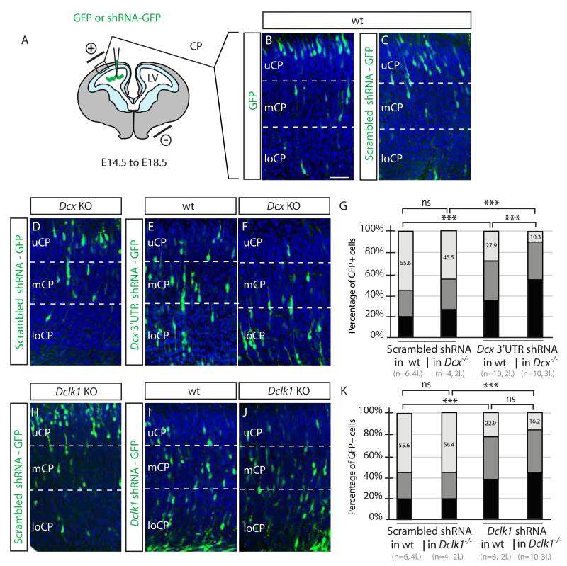
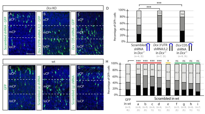
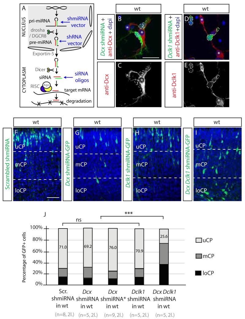
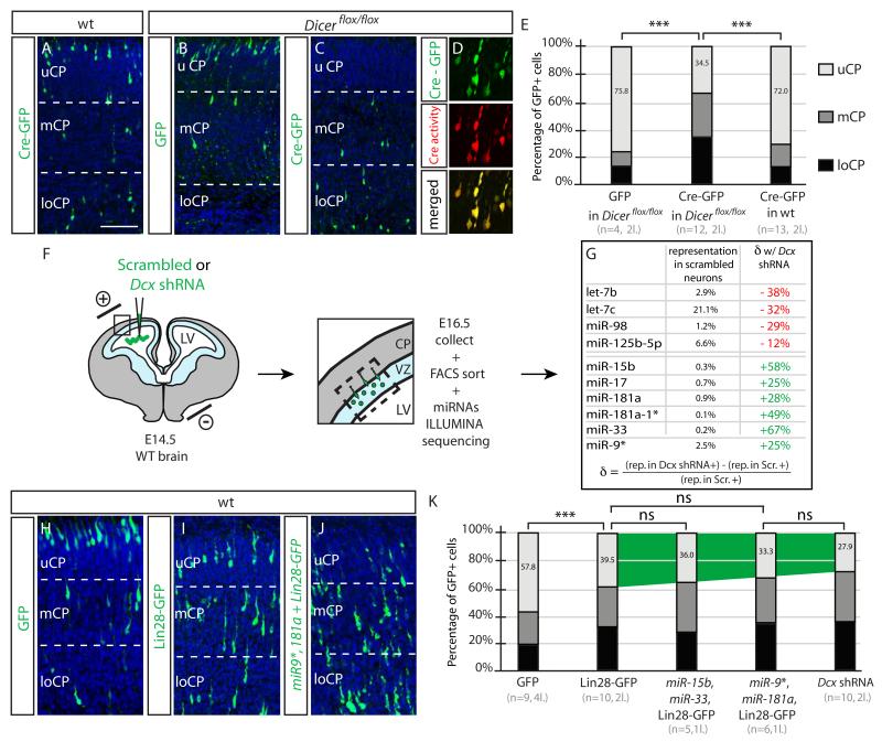
Acute gene inactivation using short hairpin RNA (shRNA, knockdown) in developing brain is a powerful technique to study genetic function; however, discrepancies between knockdown and knockout murine phenotypes have left unanswered questions. For example, doublecortin (Dcx) knockdown but not knockout shows a neocortical neuronal migration phenotype. Here we report that in utero electroporation of shRNA, but not siRNA or miRNA, to Dcx demonstrates a migration phenotype in Dcx knockouts akin to the effect in wild-type mice, suggesting shRNA-mediated off-target toxicity. This effect was not limited to Dcx, as it was observed in Dclk1 knockouts, as well as with a fraction of scrambled shRNAs, suggesting a sequence-dependent but not sequence-specific effect. Profiling RNAs from electroporated cells showed a defect in endogenous let7 miRNA levels, and disruption of let7 or Dicer recapitulated the migration defect. The results suggest that shRNA-mediated knockdown can produce untoward migration effects by altering endogenous miRNA pathways.


Figures

Figure 1
Figure 1
Knockdown vs. knockout discrepancy in Dcx and Dclk1 mice is at least partly due to off-target effects of shRNA (A-C) DNA injected into the cerebral lateral ventricle (LV) at E14.5, electroporated into the ventricular zone (light blue), then GFP+ cell positions assessed at E18.5, dividing the cortex into upper, middle and lower cortical plate regions (uCP, mCP, loCP). In WT with empty GFP or Scrambled shRNA-GFP, most labeled cells were located in uCP. Scale bar 100 um. (D-F) Scrambled shRNA into Dcx knockout (KO) showed no defect, whereas Dcx 3′UTR shRNA into either WT or Dcx KO showed a migration defect, with most cells in mCP and loCP. (G) Scrambled shRNA in WT or Dcx KO showed 55.6% vs. 45.5% of cells in UCP. There was a mild though not significant difference in Dcx KO with scrambled shRNA (ns). n = 4-6 mice from 2-4 litters for each condition. p >0.05, Student t-test. Dcx 3′UTR shRNA impaired migration in both the WT and Dcx KO, with 27.9% vs. 10.3% of cells in UCP (***, p < 0.001). (H-K) Scrambled shRNA in WT vs. Dclk1 KO compared with Dclk1 CD shRNA in WT vs. Dclk1 KO showed 55.6% vs. 56.4%, and 22.9 vs. 16.2% of cells in UCP, indicating an effect of the Dclk1 shRNA, irrespective of mouse genotype. n = 6-10 mice from 2-4 litters for each condition. See also Figure S1.
Figure 2
Figure 2
Sequence dependent but not sequence specific off-target effects of shRNAs (A-D) Dcx KO electroporated with swapped loop (blue) maintaining Dcx 3′UTR or CDS stem sequence, showed migration defects (13.9% vs. 19.0% of cells in uCP, compared with 45.5% in Scrambled, ***, p < 0.001). Scale bar 100 um. (E-H) Severe, Moderate or None migration defects induced by a scrambled shRNAs. Nine scrambled shRNAs electroporated into WT mice, ranked from most to least severe effects, compared with GFP alone. The first four (a-d) showed the most dramatic differences (a-d, 32.1-43.1% in uCP, ***, p < 0.001), the next showed moderate difference (e, 51.3% in uCP, * p < 0.01), and last four were not significant (f-i, 51.3-1.9%, ns) compared with GFP alone. n = 4-9 mice from 1-4 litters for each condition. See also Figure S2.
Figure 3
Figure 3
shmiRNA or siRNA fail to induce off-target migration defects (A) The pri-mRNA is cleaved in the nucleus by drosha/DGCR8 to yield pre-miRNA, which is akin to products from an shRNA vector, containing an antisense (red) and * (green) strand, exported through the nuclear pore (space between ovals) by Exportin-5, further cleaved in the cytoplasm by Dicer to yield siRNAs, akin to synthetic siRNA duplex oligos, loaded into the RISC complex and then guide strand associates with the target mRNA for silencing. (B-E) Dcx and Dclk1 miRNA are effective against endogenous Dcx or Dclk1 protein in transfected primary cortical neurons (green). Bar 50 um. (F-I) Dcx and Dclk1 shmiRNAs together but neither separately induces migration defect. Bar 100 um. (J) Specificity and sensitivity of shmiRNAs targeting Dcx and Dclk1in WT cortex (71.0% Scrambled vs. 69.2% Dcx shmiRNA1 vs. 76.0% Dcx shmiRNA2 vs. 70.9% Dclk1 shmiRNA vs. 25.6% Dcx;Dclk1 shmiRNA KD of cells in UCP, ***, p 0.001). n = 5-9 mice from each of 2 litters for each condition. See also Figure S3.
Figure 4
Figure 4
Disruption of endogenous miRNA processing blocks neuronal migration (A-C) Dicerflox/flox Cre-GFP electroporated neurons fail neuronal migration. (D) Co-electroporation with Cre-reporter (DsRed2 expressing). Bar 100 um. (E) Percent GFP+ cells in uCP. Defect only seen for Cre-GFP electroporation into Dicerflox/flox mice. ***, p < 0.001. n = 4-13 mice in 2 litters for each condition. (F) Following electroporation, VZ and loCP was microdissected at E16.5, cells dissociated, FACS isolated, then miRNAs sequenced. (G) miRNAs with most severely dysregulated levels either reduced (red) or increased (green). The percent representation of each miRNA relative to total miRNAs identified (>20,000 reads). Formula to calculate the change (δ) in miRNA. (H-I) WT mice electroporated with GFP alone, Lin28-GFP (to interfere with let-7 family) and combination of miR9*, 181a and Lin28-GFP, showing progressive more severe migration defects. (K) Migration defects of combination electroporations, compared with Dcx shRNA. n = 5-10 mice from each of 1-4 litters for each condition.

References

    1. Ahmed F, Ansari HR, Raghava GP. Prediction of guide strand of microRNAs from its sequence and secondary structure. BMC Bioinformatics. 2009;10:105. - PMC - PubMed
    1. Alvarez VA, Ridenour DA, Sabatini BL. Retraction of synapses and dendritic spines induced by off-target effects of RNA interference. J Neurosci. 2006;26:7820–7825. - PMC - PubMed
    1. Bai J, Ramos RL, Ackman JB, Thomas AM, Lee RV, LoTurco JJ. RNAi reveals doublecortin is required for radial migration in rat neocortex. Nat Neurosci. 2003;6:1277–1283. - PubMed
    1. Balzer E, Moss EG. Localization of the developmental timing regulator Lin28 to mRNP complexes, P-bodies and stress granules. RNA biology. 2007;4:16–25. - PubMed
    1. Bauer M, Kinkl N, Meixner A, Kremmer E, Riemenschneider M, Forstl H, Gasser T, Ueffing M. Prevention of interferon-stimulated gene expression using microRNA-designed hairpins. Gene therapy. 2009;16:142–147. - PubMed

Publication types