. 2014 Jun 19;10(6):e1004187.
doi: 10.1371/journal.ppat.1004187. eCollection 2014 Jun.

A translocated effector required for Bartonella dissemination from derma to blood safeguards migratory host cells from damage by co-translocated effectors

Affiliations

A translocated effector required for Bartonella dissemination from derma to blood safeguards migratory host cells from damage by co-translocated effectors

Rusudan Okujava et al. PLoS Pathog. .

Abstract

Numerous bacterial pathogens secrete multiple effectors to modulate host cellular functions. These effectors may interfere with each other to efficiently control the infection process. Bartonellae are Gram-negative, facultative intracellular bacteria using a VirB type IV secretion system to translocate a cocktail of Bartonella effector proteins (Beps) into host cells. Based on in vitro infection models we demonstrate here that BepE protects infected migratory cells from injurious effects triggered by BepC and is required for in vivo dissemination of bacteria from the dermal site of inoculation to blood. Human endothelial cells (HUVECs) infected with a ΔbepE mutant of B. henselae (Bhe) displayed a cell fragmentation phenotype resulting from Bep-dependent disturbance of rear edge detachment during migration. A ΔbepCE mutant did not show cell fragmentation, indicating that BepC is critical for triggering this deleterious phenotype. Complementation of ΔbepE with BepEBhe or its homologues from other Bartonella species abolished cell fragmentation. This cyto-protective activity is confined to the C-terminal Bartonella intracellular delivery (BID) domain of BepEBhe (BID2.EBhe). Ectopic expression of BID2.EBhe impeded the disruption of actin stress fibers by Rho Inhibitor 1, indicating that BepE restores normal cell migration via the RhoA signaling pathway, a major regulator of rear edge retraction. An intradermal (i.d.) model for B. tribocorum (Btr) infection in the rat reservoir host mimicking the natural route of infection by blood sucking arthropods allowed demonstrating a vital role for BepE in bacterial dissemination from derma to blood. While the Btr mutant ΔbepDE was abacteremic following i.d. inoculation, complementation with BepEBtr, BepEBhe or BIDs.EBhe restored bacteremia. Given that we observed a similar protective effect of BepEBhe on infected bone marrow-derived dendritic cells migrating through a monolayer of lymphatic endothelial cells we propose that infected dermal dendritic cells may be involved in disseminating Bartonella towards the blood stream in a BepE-dependent manner.


Conflict of interest statement

The authors have declared that no competing interests exist.

Figures
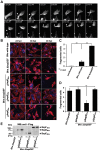
Figure 1
Figure 1. Infection of HUVECs with the Bhe ΔbepDEF mutant leads to cell fragmentation.
Subconfluent monolayers of HUVECs were infected with the indicated bacterial strains at a MOI = 200. (A) HUVECs expressing LifeAct-mCherry were infected with Bhe ΔbepDEF and subjected to live cell imaging with an MD ImageXpress Micro automated microscope. Snapshots of gray scale images taken at different time points as depicted by the time stamps (format: dd: hh:mm) are presented (scale bar = 50 µm). The arrowheads are pointing to the regions of the cell where the fragmentation is taking place. (B) HUVECs infected with the indicated bacterial strains were fixed at 24 h, 36 h and 48 h post infection followed by immunocytochemical staining and confocal laser scanning microscopy. F-actin is represented in red and DNA in blue (scale bar = 50 µm). (C, D) Quantification of cell fragmentation at 48 hpi was performed in semi-automated manner. Images were acquired in 96 well-plate format by MD ImageXpress Micro automated microscopes with 10× magnification. The number of fragmented cells (cells with thin and multipolar elongations) were defined by eye and counted manually. The percentage of fragmented cells is normalized to Bhe wild-type infection. In each condition triplicate wells with each 10 randomly picked fields were imaged and presented as mean +/− SD. Statistical significance was determined using Student's t-test. P<0.05 was considered statistically significant. Data from one representative experiment (n = 3) are presented. (E) Protein levels of the BepDBhe, BepEBhe and BepFBhe by plasmid overexpression in ΔbepDEF. The anti-Flag western blot was obtained from total lysate of corresponding Bhe strains.
Figure 2
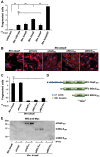
Figure 2. Deletion of BepE is sufficient for Bhe to induce cell fragmentation.
(A–C) Subconfluent monolayers of HUVECs were infected with MOI = 200 of the indicated bacterial strains. (B) Infected HUVECs were fixed at 48 h post infection followed by immunocytochemical staining and confocal laser scanning microscopy. F-actin is represented in red and DNA in blue (scale bar = 50 µm). (A) and (C) quantification of cell fragmentation at 48 h post infection was performed as described for Fig. 1C and D. The mean and SD of triplicate samples is presented. Statistical significance was determined using Student's t-test. P<0.05 was considered statistically significant. Data from one representative experiment (n = 3) are presented. (D) Schematic view of BepEBhe and N-terminal deletion mutants expressed in Bartonella from a plasmid. (E) Protein levels of the BepEBhe mutants shown in figure by overexpression in Bhe ΔbepE. The anti-Myc western blot was obtained from total lysate of corresponding Bartonella strains.
Figure 3
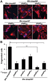
Figure 3. Expression of BepEBhe homologues in the Bhe ΔbepDEF inhibit the cell fragmentation phenotype.
(A) Subconfluent monolayers of HUVECs were infected for 48 h with MOI = 200 of the Bhe ΔbepDEF mutant complemented with the indicated Bep-expression plasmids followed by fixation, immunocytochemical staining and confocal laser scanning microscopy. F-actin is represented in red and DNA in blue (scale bar = 50 µm). (B) Quantification of cell fragmentation at 48 h post infection was performed as described for Fig. 1C and D and presented as mean of triplicate samples +/− SD. Statistical significance was determined using Student's t-test. P<0.05 was considered statistically significant. Data from one representative experiment (n = 3) are presented.
Figure 4
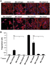
Figure 4. The double deletion mutant Bhe ΔbepCE abolishes cell fragmentation.
(A) Subconfluent monolayers of HUVECs were infected for 48 h with MOI = 200 of the Bhe strains depicted in the figure or were left uninfected. Samples were then fixed, stained immunocytochemically and analyzed by confocal laser scanning microscopy. F-actin is represented in red (Phalloidin) and DNA in blue (DAPI) (scale bar = 50 µm). (B) Quantification of cell fragmentation at 48 h post infection was performed as described for Fig. 1C and D and presented as mean of triplicate samples +/− SD. Statistical significance was determined using Student's t-test. P<0.05 was considered statistically significant. Data from one representative experiment (n = 3) are presented.
Figure 5
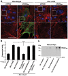
Figure 5. BepE protects host cells from fragmentation upon translocation via T4SS.
(A) Subconfluent monolayers of HUVECs were infected with MOI = 100 of the indicated bacterial strains for 24 h. After fixation and subsequent immunocytochemical staining the specimen was analyzed by confocal laser scanning microscopy. F-actin is represented in red (Phalloidin) and DNA in Blue (DAPI). Translocation of the effector protein into the infected cells was detected by anti-Myc-staining depicted in green (scale bar = 20 µm). (B) Subconfluent monolayers of HUVECs were infected with MOI = 200 or MOI = 200+200 in case of mixed infection depicted in the figure. Quantification of cell fragmentation at 48 h post infection was performed as described for Fig. 1C and D and presented as mean of triplicate samples +/− SD. Statistical significance was determined using Student's t-test. P<0.05 was considered statistically significant. Data from one representative experiment (n = 2) are presented. (C) Protein levels of the BepEBhe by overexpression in Bartonella strains. The anti-Myc western blot was obtained from total lysate of corresponding Bartonella strains depicted in figure.
Figure 6
Figure 6. Ectopic expression of BepEBhe in HUVECs prevents cell fragmentation.
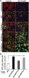
(A, B) HUVECs of an early passage were transduced with lentiviruses for the expression of the depicted GFP-fusion proteins. The mixed culture of transduced and non-transduced cells were infected with the indicated Bhe strains (MOI = 200). Infected cells were either fixed and stained for microscopy or analyzed for the survival by FACS at 48 hpi. (A) Representative microscopy images (scale bar = 100 µm). F-actin is represented in red (Phalloidin), DNA in blue (DAPI), GFP in green. (B) Protection by GFP-fused BepE and its derivatives against fragmentation induced by Bhe ΔbepDEF mutant strains. GFP-positive cell were quantified by FACS and normalized to the uninfected cell population. One representative experiment (n = 3) with the mean of triplicate samples +/− SD are presented. Statistical significance was determined using Student's t-test. P<0.05 was considered statistically significant.
Figure 7
Figure 7. BepEBhe localizes to cell-to-cell contacts and is recruited to the plasma membrane of HUVECs following translocation via the T4SS or by ectopic expression.
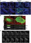
(A) Subconfluent monolayers of HUVECs were infected with MOI = 100 of the indicated bacterial strains for 24 h or left uninfected. After fixation and subsequent immunocytochemical staining the specimen was analyzed by confocal laser scanning microscopy. F-actin is represented in blue (Phalloidin) and VE-cadherin staining in red (anti-VE-cadherin). Translocation of the effector protein into the infected cells was detected by anti Myc-staining depicted in green (scale bar = 20 µm). (B) HUVECs of an early passage were transduced with lentiviruses directing expression of either GFP or GFP-BepEBhe. Cells were stained with wheat germ agglutinin (WGA, red) and fixed. Confocal images were acquired in xy- and xz-planes (scale bar = 50 µm). (C) gfp-bepEBhe-transduced HUVECs were subjected to live cell imaging using an MD ImageXpress Micro automated microscope. Snapshots of gray scale images at different time points as depicted by the time stamps (format: dd:hh:mm) are presented (scale bar = 50 µm). The arrows are pointing to the regions of transient enrichments of BepEBhe in migrating HUVECs.
Figure 8
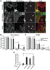
Figure 8. BepEBhe interferes with the effect of Rho inhibitor I.
(A) A mixed population of HUVECs lentivirally transduced with a GFP-BepEBhe construct (green) were treated either with 0.5 µg/ml Rho inhibitor I or 10 µM Y27632 (4 h and 30 min respectively) or were left untreated. Cells were fixed, followed by staining for F-actin with Phalloidin (red) and nucleus with DAPI (blue). Representative confocal images are depicted which show selective retainment of actin stress fibers in the GFP-BepEBhe-expressing subpopulation (scale bar = 50 µm). (B) Dose-dependent effect of Rho inhibitor I on HUVECs expressing GFP-BepEBhe. Cells with actin stress fibers in GFP- or GFP-BepEBhe-transduced or non-transduced HUVECs were quantified in semi-automated manner, similarly as described for Fig. 1C and D. The percentage of stress fiber-containing cells is shown in the figure. In each condition triplicate wells with each 10 randomly picked microscopic fields were analyzed and represented as mean +/− SD. Statistical significance was determined using Student's t-test. P<0.05 was considered statistically significant. Data from one representative experiment (n = 3) are presented. (C) Dose-dependent effect of Y27632 on HUVECs expressing GFP-BepEBhe. Cells with actin stress fibers within GFP or GFP-BepEBhe-transduced or non-transduced HUVECs were quantified in semi-automated manner as described for (B). (D) Comparison of the potency of GFP-BepEBhe and mutant derivatives to interfere with Rho inhibitor I. Lentivirally transduced HUVECs expressing GFP-fusions of BepEBhe and the depicted mutant derivatives were treated with 0.5 µg/ml Rho inhibitor I. Specimen were stained and stress fiber-containing cells were quantified as described in (B).
Figure 9
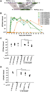
Figure 9. BepE is essential for Bartonella tribocorum (Btr) to establish bacteremia after intradermal (i.d.) infection of the rat reservoir host.
(A) Domain organization of BepE orthologues in Btr and Bhe. The BepE homologues from Bartonella species depicted in the figure (BepEBhe, BepDBtr, BepEBtr) were aligned using Geneious Pro 5.3.4. The amino acid sequence alignment with pairwise % identity is indicated. The tyrosine-containing N-termini and BID domains were aligned independently. (B) Btr ΔbepDE is not able to reach the blood of rats infected by the i.d. route. Rats (n = 5) were inoculated in the ear dermis with either Btr wild-type or Btr ΔbepDE. Blood was drawn at the indicated days post infection (dpi), diluted and plated on sheep blood supplemented Columbia agar plates (CBA) for counting of colony forming units (CFUs). (C) Complementation of the Btr ΔbepDE mutant with BepE is sufficient to restore bacteremia in rats infected by the i.d. route. Groups of rats (n≥3) were infected with the indicated strains by the i.v. or i.d. route. Blood was drawn at 16 dpi and CFUs were recovered as described for B. The graph represents CFUs/ml of blood for individual animals (circles) and their cohort mean (line). Statistical significance was determined using Student's t-test. P<0.05 was considered statistically significant. (D) Heterologous complementation of Btr ΔbepDE with pBIDs.EBhe is sufficient to rescue the abacteremia phenotype following infection by the i.d. route. The infections were performed as described for (C). Data represented for BIDs.EBhe complementation were acquired in separate experiment from the other data shown. P<0.05 was considered statistically significant.
Figure 10
Figure 10. Dendritic cells are infected by Bartonella.
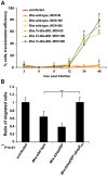
(A) Effector translocation by Bhe into mouse bone marrow-derived dendritic cells (BMDCs). Balb/c mouse BMDCs were infected with corresponding MOIs and strains. “Effector”, Bla-BID, translocation efficiency was assessed as the % of infected cells that converted CCF2-AM blue emission into green detected by Leica DM-IRBE inverted fluorescence microscope. The bars represent the mean of triplicate samples +/− SD. Data from one representative experiment (n = 2) are presented. (B) Migration of BMDCs is inhibited in a trans-well assay by Bhe ΔbepDEF infection. BMDCs were pre-infected with MOI = 50 of the indicated bacterial strains. Infected cells were embedded in collagen and mounted in a trans-well migration system that was prior seeded with a confluent monolayer of iLECs (immortalized lymphatic endothelial cells). BMDCs that migrated though the iLECs were quantified after 24 h. The data normalized to uninfected condition. The bars represent the mean of triplicate samples +/− SD. Statistical significance was determined using Student's t-test. P<0.05 was considered statistically significant. Data from one representative experiment (n = 3) are presented.

Comment in

Similar articles

Cited by

References

    1. Monack DM, Mueller A, Falkow S (2004) Persistent bacterial infections: the interface of the pathogen and the host immune system. Nat Rev Microbiol 2: 747–765. - PubMed
    1. Lee KS, Kalantzis A, Jackson CB, O'Connor L, Murata-Kamiya N, et al. (2012) Helicobacter pylori CagA triggers expression of the bactericidal lectin REG3gamma via gastric STAT3 activation. PLoS One 7: e30786. - PMC - PubMed
    1. Kido M, Watanabe N, Aoki N, Iwamoto S, Nishiura H, et al. (2011) Dual roles of CagA protein in Helicobacterpylori-induced chronic gastritis in mice. Biochem Biophys Res Commun 412: 266–272. - PubMed
    1. Schroeder GN, Hilbi H (2008) Molecular pathogenesis of Shigella spp.: controlling host cell signaling, invasion, and death by type III secretion. Clin Microbiol Rev 21: 134–156. - PMC - PubMed
    1. Diacovich L, Gorvel JP (2010) Bacterial manipulation of innate immunity to promote infection. Nat Rev Microbiol 8: 117–128. - PubMed

Publication types

MeSH terms

LinkOut - more resources