

doi: 10.1371/journal.ppat.1003983.
eCollection 2014 Jun.
Antibody responses to Mycoplasma pneumoniae: role in pathogenesis and diagnosis of encephalitis?
Affiliations
- PMID: 24945969
- PMCID: PMC4055762
- DOI: 10.1371/journal.ppat.1003983
Item in Clipboard
Antibody responses to Mycoplasma pneumoniae: role in pathogenesis and diagnosis of encephalitis?
PLoS Pathog.
.
No abstract available
Conflict of interest statement
The authors have declared that no competing interests exist.
Figures
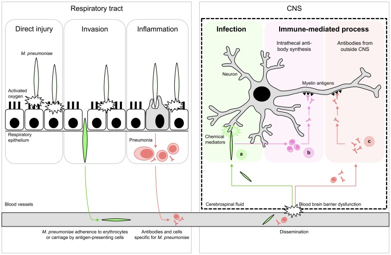
(Left) Respiratory tract infection. M. pneumoniae resides mostly extracellularly on epithelial surfaces. Its close association allows the production of direct injury by a variety of local cytotoxic effects. Furthermore, it can induce inflammatory responses, elicited by both adhesion proteins and glycolipid epitopes that result in pneumonia. (Right) Encephalitis. Extrapulmonary disease of the CNS is characterized by systemic dissemination with resultant direct infection and local tissue injury (A) or immune-mediated injury (B,C). The latter may occur as a result of cross-reactive antibodies against myelin components, e.g., gangliosides and galactocerebroside C. These antibodies could theoretically have originated from intrathecal synthesis (B) or from outside the CNS (C). Figure adapted from ; see references in the text.
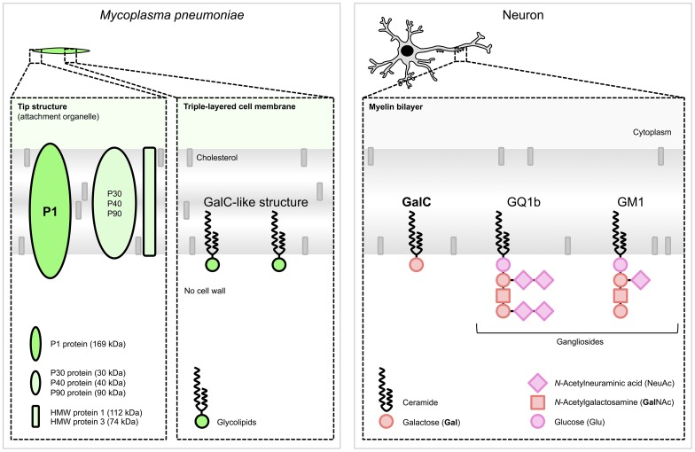
(Left) M. pneumoniae adhesion proteins and glycolipids. The immunogenic and major cytadherence proteins P1 and P30 are densely clustered at the tip structure. The P1 protein and glycolipids, e.g., those forming a GalC-like structure , elicit cross-reactive antibodies induced by molecular mimicry. (Right) Host myelin glycolipids, to which antibodies were found in patients with M. pneumoniae encephalitis. Glycolipids are organized in specialized functional microdomains called “lipid rafts” and play a part in the maintenance of the cell membrane structure. Abbreviations: GalC, galactocerebroside C; GQ1b, ganglioside quadrosialo 1b; GM1, ganglioside monosialo 1 (the numbers stand for the order of migration on thin-layer chromatography, and the lower-case letters stand for variations within basic structures); HMW, high-molecular-weight. Structures of M. pneumoniae adhesion proteins and host glycolipids are adapted from and , respectively.
References
-
- Narita M (2009) Pathogenesis of neurologic manifestations of Mycoplasma pneumoniae infection. Pediatr Neurol 41: 159–166. - PubMed
-
- Foy HM (1993) Infections caused by Mycoplasma pneumoniae and possible carrier state in different populations of patients. Clin Infect Dis 17 Suppl 1 S37–S46. - PubMed
-
- Lind K, Benzon MW, Jensen JS, Clyde WA Jr (1997) A seroepidemiological study of Mycoplasma pneumoniae infections in Denmark over the 50-year period 1946–1995. Eur J Epidemiol 13: 581–586. - PubMed
-
- Michelow IC, Olsen K, Lozano J, Rollins NK, Duffy LB, et al. (2004) Epidemiology and clinical characteristics of community-acquired pneumonia in hospitalized children. Pediatrics 113: 701–707. - PubMed
-
- Baer G, Engelcke G, Abele-Horn M, Schaad UB, Heininger U (2003) Role of Chlamydia pneumoniae and Mycoplasma pneumoniae as causative agents of community-acquired pneumonia in hospitalised children and adolescents. Eur J Clin Microbiol Infect Dis 22: 742–745. - PubMed
Publication types
MeSH terms
Substances
LinkOut - more resources
Full Text Sources
Other Literature Sources
Medical
