Review
. 2014 Jul;24(4):387-95.
doi: 10.1111/bpa.12158.

PDGF, pericytes and the pathogenesis of idiopathic basal ganglia calcification (IBGC)

Affiliations
Review

PDGF, pericytes and the pathogenesis of idiopathic basal ganglia calcification (IBGC)

Christer Betsholtz et al. Brain Pathol. 2014 Jul.

Abstract

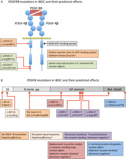
Platelet-derived growth factors (PDGFs) are important mitogens for various types of mesenchymal cells, and as such, they exert critical functions during organogenesis in mammalian embryonic and early postnatal development. Increased or ectopic PDGF activity may also cause or contribute to diseases such as cancer and tissue fibrosis. Until recently, no loss-of-function (LOF) mutations in PDGF or PDGF receptor genes were reported as causally linked to a human disease. This changed in 2013 when reports appeared on presumed LOF mutations in the genes encoding PDGF-B and its receptor PDGF receptor-beta (PDGF-Rβ) in familial idiopathic basal ganglia calcification (IBGC), a brain disease characterized by anatomically localized calcifications in or near the blood microvessels. Here, we review PDGF-B and PDGF-Rβ biology with special reference to their functions in brain-blood vessel development, pericyte recruitment and the regulation of the blood-brain barrier. We also discuss various scenarios for IBGC pathogenesis suggested by observations in patients and genetically engineered animal models of the disease.

Keywords: Pericytes.


Figures

Figure 1
Figure 1
Schematic representation of the mutations reported to date in PDGFB and PDGFRB and their predicted effects on protein function. A. A schematic representation of PDGFRβ with PDGFBB bound to the extracellular ligand‐binding domain. Circles in PDGFRβ indicate immunoglobulin domains and boxes represent kinase domains. The three idiopathic basal ganglia calcification (IBGC) mutations reported to date are indicated by type and approximate position. Note that one of the mutations is predicted to be damaging to the receptor functions, as it introduces a proline residue close to the ATP‐binding pocket of the receptor kinase. Proline residues are known to have strong effects on the three‐dimensional (3D) structure in this region. B. The PDGFB precursor protein is indicated in different shades of gray. SS, signal sequence for secretion; N‐term. PP, amino‐terminal pro‐peptide; GF domain, growth factor domain; Ret. Motif, heparan sulfate proteoglycan‐binding extracellular matrix retention motif. The nature and approximate position of the mutations are indicated and their effects described in boxes with the same color. Haploinsufficiency indicates that mere loss of one functional copy of the PDGFB gene is disease causing. For the mutants with putative residual cysteine residues engaged in ligand dimerization, a dominant‐negative effect might be conceived, which in theory predicts a 75% reduction of the amount produced functional PDGFBB.
Figure 2
Figure 2
Cartoon illustrating the changes at the neurovascular unit (NVU) in idiopathic basal ganglia calcification (IBGC). The illustration takes into account observations made in genetically engineered mouse models of IBGC. A. Cross section of a normal NVU. Endothelial cells (E) are surrounded by a basement membrane (BM), pericytes (P) and astrocyte end‐feet (AEF). Microglia (M) make contacts to the NVU through cytoplasmic processes. B. Altered features of the NVU in IBGC. Calcified nodules are associated with blood vessels and are surrounded by reactive astrocytes and microglia. A hypothetical bone‐forming cell (red) initiates and propagates the formation and growth of the calcified nodule. The origin of the bone‐forming cell is unclear, but available literature provides precedence for several possible origins, as indicated (dashed line with question mark). Mouse models of IBGC provide a correlation between pericyte deficiency and BBB disruption, on the one hand, and brain calcification, on the other hand. The altered properties of endothelial cells and pericytes are indicated by their changed shape.

References

    1. Abramsson A, Kurup S, Busse M, Yamada S, Lindblom P, Schallmeiner E et al (2007) Defective N‐sulfation of heparan sulfate proteoglycans limits PDGF‐BB binding and pericyte recruitment in vascular development. Genes Dev 21:316–331. - PMC - PubMed
    1. Abramsson A, Lindblom P, Betsholtz C (2003) Endothelial and nonendothelial sources of PDGF‐B regulate pericyte recruitment and influence vascular pattern formation in tumors. J Clin Invest 112:1142–1151. - PMC - PubMed
    1. Andrae J, Gallini R, Betsholtz C (2008) Role of platelet‐derived growth factors in physiology and medicine. Genes Dev 22:1276–1312. - PMC - PubMed
    1. Armulik A, Abramsson A, Betsholtz C (2005) Endothelial/pericyte interactions. Circ Res 97:512–523. - PubMed
    1. Armulik A, Genové G, Betsholtz C (2011) Pericytes: developmental, physiological, and pathological perspectives, problems, and promises. Dev Cell 21:193–215. - PubMed

Publication types

MeSH terms

Substances

Supplementary concepts