. 2014 Jul;24(4):396-403.
doi: 10.1111/bpa.12159.

Failure of perivascular drainage of β-amyloid in cerebral amyloid angiopathy

Affiliations

Failure of perivascular drainage of β-amyloid in cerebral amyloid angiopathy

Cheryl A Hawkes et al. Brain Pathol. 2014 Jul.

Abstract

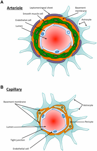
In Alzheimer's disease, amyloid-β (Aβ) accumulates as insoluble plaques in the brain and deposits in blood vessel walls as cerebral amyloid angiopathy (CAA). The severity of CAA correlates with the degree of cognitive decline in dementia. The distribution of Aβ in the walls of capillaries and arteries in CAA suggests that Aβ is deposited in the perivascular pathways by which interstitial fluid drains from the brain. Soluble Aβ from the extracellular spaces of gray matter enters the basement membranes of capillaries and drains along the arterial basement membranes that surround smooth muscle cells toward the leptomeningeal arteries. The motive force for perivascular drainage is derived from arterial pulsations combined with the valve effect of proteins present in the arterial basement membranes. Physical and biochemical changes associated with arteriosclerosis, aging and possession of apolipoprotein E4 genotype lead to a failure of perivascular drainage of soluble proteins, including Aβ. Perivascular cells associated with arteries and the lymphocytes recruited in the perivenous spaces contribute to the clearance of Aβ. The failure of perivascular clearance of Aβ may be a major factor in the accumulation of Aβ in CAA and may have significant implications for the design of therapeutics for the treatment of Alzheimer's disease.

Keywords: Alzheimer's disease; amyloid β; cerebral amyloid angiopathy; interstitial fluid neuroimmunology; perivascular drainage.


Figures

Figure 1
Figure 1
A. Diagrammatic representation of a cross section through a cerebral cortical arteriole. There are no elastic laminae, but there is a tunica media with smooth muscle cells (green) and basement membrane (orange) interposed between the smooth muscle cells. The astrocyte end‐feet (light blue) abut upon the leptomeningeal sheet that reflects upon the artery as it penetrates the brain from the subarachnoid space. B. Diagrammatic representation of a capillary in the cerebral cortex. The endothelial cells are connected by tight junctions. On the abluminal border of the endothelial cells there is a basement membrane that results from the fusion of the basement membranes of the endothelium and the astrocyte end‐feet. Pericytes are completely surrounded by basement membrane.
Figure 2
Figure 2
A. Diagrammatic representation of perivascular clearance of solutes, normally entering the walls (basement membranes) of capillaries and then arteries, but not of veins. B. Cross section through a human artery at the surface of the brain. The tissue has been immunostained for collagen IV (a marker of basement membranes, shown here in blue), smooth muscle cells (immunostaining for smooth muscle actin, shown in green) and amyloid‐β (Aβ), using anti‐4G8 antibody, shown here in red. Note the presence of Aβ surrounding the smooth muscle cells, leaving the endothelial basement membrane (blue) free of Aβ. There is some Aβ accumulating at the surface of the brain, with the collagen IV present in glia limitans. Confocal microscopy. Scale bar: 20 μm.

References

    1. Agrawal S, Anderson P, Durbeej M, van Rooijen N, Ivars F, Opdenakker G, Sorokin LM (2006) Dystroglycan is selectively cleaved at the parenchymal basement membrane at sites of leukocyte extravasation in experimental autoimmune encephalomyelitis. J Exp Med 203:1007–1019. - PMC - PubMed
    1. Arbel‐Ornath M, Hudry E, Eikermann‐Haerter K, Hou S, Gregory JL, Zhao L et al (2013) Interstitial fluid drainage is impaired in ischemic stroke and Alzheimer's disease mouse models. Acta Neuropathol 126:353–364. - PMC - PubMed
    1. Ashford JW (2004) APOE genotype effects on Alzheimer's disease onset and epidemiology. J Mol Neurosci 23:157–165. - PubMed
    1. Ball KK, Cruz NF, Mrak RE, Dienel GA (2010) Trafficking of glucose, lactate, and amyloid‐beta from the inferior colliculus through perivascular routes. J Cereb Blood Flow Metab 30:162–176. - PMC - PubMed
    1. Barua NU, Bienemann AS, Hesketh S, Wyatt MJ, Castrique E, Love S, Gill SS (2012) Intrastriatal convection‐enhanced delivery results in widespread perivascular distribution in a pre‐clinical model. Fluids Barriers CNS 9:2. - PMC - PubMed

Publication types

MeSH terms