. 2014 Jul;24(4):404-13.
doi: 10.1111/bpa.12162.

The fibrotic scar in neurological disorders

Affiliations

The fibrotic scar in neurological disorders

Francisco Fernández-Klett et al. Brain Pathol. 2014 Jul.

Abstract

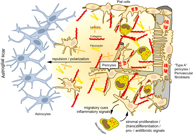
Tissue fibrosis, or scar formation, is a common response to damage in most organs of the body. The central nervous system (CNS) is special in that fibrogenic cells are restricted to vascular and meningeal niches. However, disruption of the blood-brain barrier and inflammation can unleash stromal cells and trigger scar formation. Astroglia segregate from the inflammatory lesion core, and the so-called "glial scar" composed of hypertrophic astrocytes seals off the intact neural tissue from damage. In the lesion core, a second type of "fibrotic scar" develops, which is sensitive to inflammatory mediators. Genetic fate mapping studies suggest that pericytes and perivascular fibroblasts are activated, but other precursor cells may also be involved in generating a transient fibrous extracellular matrix in the CNS. The stromal cells sense inflammation and attract immune cells, which in turn drive myofibroblast transdifferentiation. We believe that the fibrotic scar represents a major barrier to CNS regeneration. Targeting of fibrosis may therefore prove to be a valuable therapeutic strategy for neurological disorders such as stroke, spinal cord injury and multiple sclerosis.

Keywords: (myo)fibroblasts; extracellular matrix; glia; neuroinflammation; pericytes; platelet-derived growth factor receptor.


Figures

Figure 1
Figure 1
Cellular and molecular interactions in central nervous system (CNS) scar formation. The fibrotic scar is characterized by the deposition of extracellular matrix molecules that are otherwise scarcely expressed in the neural parenchyma such as collagens, laminins and fibronectin. These molecules are generated by stromal cells (myofibroblasts), which are normally absent from the CNS parenchyma. The stromal cells may originate from meningeal precursors (eg, pial cells of the leptomeningeal lining), perivascular fibroblasts or pericytes (“type A pericytes”). Blood‐borne macrophages and microglia contribute to the proliferation and differentiation of stromal cells by producing profibrotic mediators, and they are also involved in the resolution of the fibrous scar. Conversely, stromal cells modulate neuroinflammation by producing cytokines, chemokines and adhesion molecules. The astroglial scar is neatly separated from the fibrotic scar. Cellular and molecular constituents of the fibrotic scar induce the repulsion and polarization of astrocytes, and a new glia limitans is generated at the interphase of both cell populations.
Figure 2
Figure 2
Mural cells of the central nervous system (CNS) vasculature. This figure demonstrates the different phenotypes of mural cells along different segments of the CNS vasculature. The mural cells can be identified based on their expression of the green fluorescent protein (GFP) in a mouse line where the pericyte‐specific rgs5 gene has been replaced with a GFP reporter 56. The images represent maximal projections or three‐dimensional (3D) renderings of confocal stacks obtained from GFP‐expressing cells in the brains of rgs5 GFP/ GFP mutant mice. Nuclei are counterstained with 4′,6‐diamidino‐2‐phenylindole (DAPI). Scale bars: 10 μm. A. In a precapillary arteriole, mural cells (arrow) exhibit robust and densely packed circumferential processes, suggesting a contractile phenotype. An abluminal GFP‐expressing cell is marked by an asterisk. B. Pericytes investing capillaries (white arrows in the left panel) show a typical protruding fusiform cell body with long primary processes running in the length of the capillary. The primary processes give rise to perpendicular secondary processes. The 3D reconstruction (right panel) of the volume marked by the white box demonstrates circumferential processes underneath the pericyte cell body that surround the capillary lumen (indicated by the yellow arrow). C. Mural cells (arrow) in a postcapillary venule show a flattened, stellate morphology and loosely cover the length of the vessel.
Figure 3
Figure 3
Fibrotic and glial scar after experimental stroke. Laser confocal microscopic images of immunostained mouse brain sections at 7 days after transient middle cerebral artery occlusion 25. Nuclei are counterstained with 4′,6‐diamidino‐2‐phenylindole (DAPI). Scale bars: (a) 100 μm, (b) 20 μm. A. The infarct core exhibits an abundant infiltrate of platelet‐derived growth factor receptor (PDGFR)‐β‐immunoreactive cells (green), which is surrounded by the glial fibrillary acidic protein (GFAP)‐immunoreactive astroglial scar (red). A high‐power magnification of the area marked by the white square is shown on the right and shows the inflammatory cell infiltrate. B. PDGFR‐β‐immunoreactive cells (green) detach from CD31‐immunoreactive endothelial cells, express α‐smooth muscle actin (α‐SMA) and fibronectin (FN), and juxtapose laminin‐immunoreactive extracellular matrix (ECM) deposits (all in red).
Figure 4
Figure 4
Fibrosis in human neurological disorders. Conventional and laser confocal microscopic images of immunostained human post‐mortem brain sections. Nuclei are counterstained with hematoxylin or 4′,6‐diamidino‐2‐phenylindole (DAPI). Scale bars: 100 μm. A. Platelet‐derived growth factor receptor (PDGFR)‐β‐immunoreactive cells (brown) in a subacute stroke lesion. PDGFR‐β‐immunoreactive cells are associated with vessels in the non‐ischemic tissue (arrows), but detach from the vasculature and spread into the parenchyma (arrowheads) in the ischemic tissue (right of the white dashed line). B. PDGFR‐β‐immunoreactive cells in Alzheimer's disease (left and middle panels). PDGFR‐β‐immunoreactive cells (green) remain at perivascular sites. There is no apparent proliferation of PDGFR‐β‐immunoreactive cells around amyloid plaques (red). In addition, no ectopic deposition of laminin (green) can be detected around plaques (red) in the Alzheimer's disease brain (right panel).

References

    1. Abnet K, Fawcett JW, Dunnett SB (1991) Interactions between meningeal cells and astrocytes in vivo and in vitro . Brain Res Dev Brain Res 59:187–196. - PubMed
    1. Aldrich A, Kielian T (2011) Central nervous system fibrosis is associated with fibrocyte‐like infiltrates. Am J Pathol 179:2952–2962. - PMC - PubMed
    1. Armulik A, Genové G, Betsholtz C (2011) Pericytes: developmental, physiological, and pathological perspectives, problems, and promises. Dev Cell 21:193–215. - PubMed
    1. Armulik A, Genové G, Mäe M, Nisancioglu MH, Wallgard E, Niaudet C et al (2010) Pericytes regulate the blood‐brain barrier. Nature 468:557–561. - PubMed
    1. Asahina K, Zhou B, Pu WT, Tsukamoto H (2011) Septum transversum‐derived mesothelium gives rise to hepatic stellate cells and perivascular mesenchymal cells in developing mouse liver. Hepatology 53:983–995. - PMC - PubMed

Publication types

MeSH terms