. 2014 Jul 16;25(7):1203-12.
doi: 10.1021/bc500240p. Epub 2014 Jul 2.

Engineering protein farnesyltransferase for enzymatic protein labeling applications

Affiliations

Engineering protein farnesyltransferase for enzymatic protein labeling applications

Jonathan K Dozier et al. Bioconjug Chem. .

Abstract

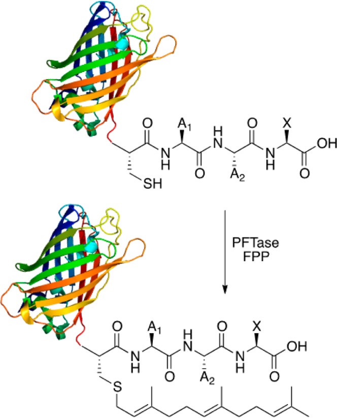
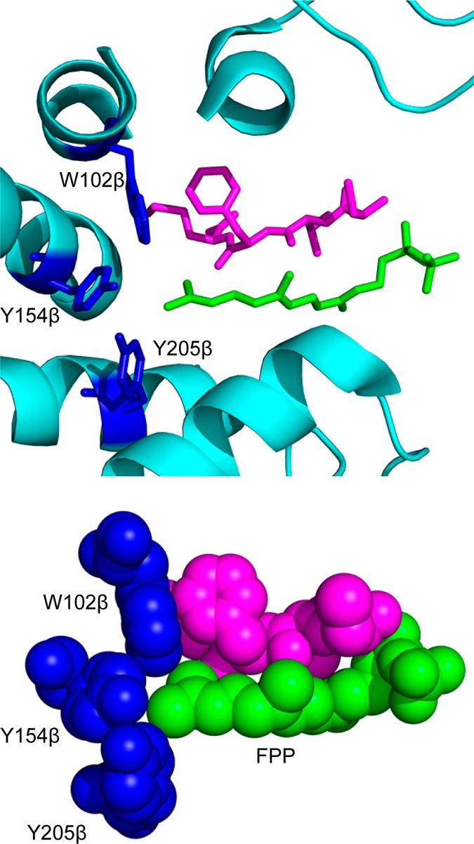
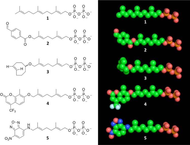
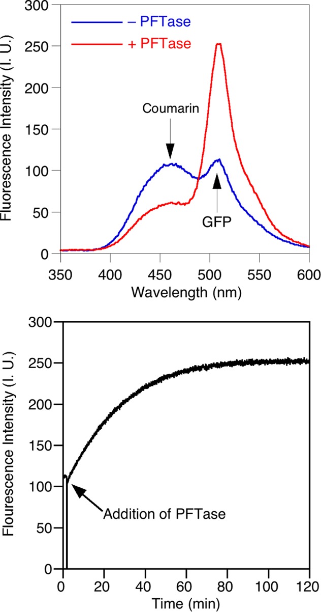
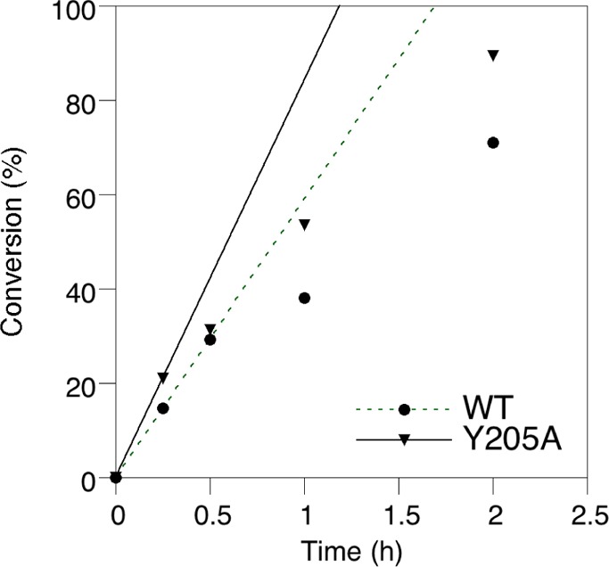
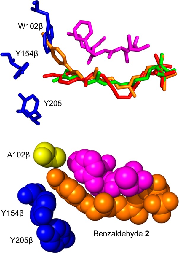
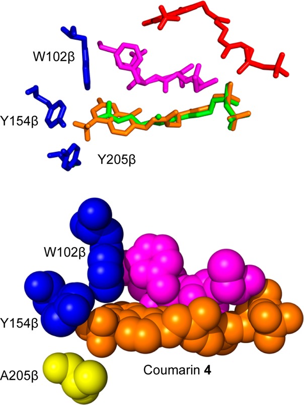
Creating covalent protein conjugates is an active area of research due to the wide range of uses for protein conjugates spanning everything from biological studies to protein therapeutics. Protein Farnesyltransferase (PFTase) has been used for the creation of site-specific protein conjugates, and a number of PFTase substrates have been developed to facilitate that work. PFTase is an effective catalyst for protein modification because it transfers Farnesyl diphosphate (FPP) analogues to protein substrates on a cysteine four residues from the C-terminus. While much work has been done to synthesize various FPP analogues, there are few reports investigating how mutations in PFTase alter the kinetics with these unnatural analogues. Herein we examined how different mutations within the PFTase active site alter the kinetics of the PFTase reaction with a series of large FPP analogues. We found that mutating either a single tryptophan or tyrosine residue to alanine results in greatly improved catalytic parameters, particularly in kcat. Mutation of tryptophan 102β to alanine caused a 4-fold increase in kcat and a 10-fold decrease in KM for a benzaldehyde-containing FPP analogue resulting in an overall 40-fold increase in catalytic efficiency. Similarly, mutation of tyrosine 205β to alanine caused a 25-fold increase in kcat and a 10-fold decrease in KM for a coumarin-containing analogue leading to a 300-fold increase in catalytic efficiency. Smaller but significant changes in catalytic parameters were also obtained for cyclo-octene- and NBD-containing FPP analogues. The latter compound was used to create a fluorescently labeled form of Ciliary Neurotrophic Factor (CNTF), a protein of therapeutic importance. Additionally, computational modeling was performed to study how the large non-natural isoprenoid analogues can fit into the active sites enlarged via mutagenesis. Overall, these results demonstrate that PFTase can be improved via mutagenesis in ways that will be useful for protein engineering and the creation of site-specific protein conjugates.


Figures

Figure 1
Figure 1
Scheme of protein prenylation. The terminal four amino acid sequence (CaaX) serves as the recognition site for the enzyme which then catalyzes the attachment of the farnesyl moiety onto the cysteine residue.
Figure 2
Figure 2
FPP binding pocket of PFTase (1JCR). (Above) The FPP binding pocket of PFTase. The isoprenoid and key residues are shown in stick representations with the protein secondary structure given in a cartoon form. Color scheme: the isoprenoid portion of FPP is shown in green. A peptide inhibitor CVFM that is bound in the CaaX substrate binding pocket is shown in purple. Key amino acid residues in the binding pocket are shown in blue. (Below) Space filling representation of the PFTase binding pocket. The color scheme is the same as that used in the top panel.
Figure 3
Figure 3
Structures of farensyl diphosphate and analogues. (Left) Lewis structure representation. (Right) Extended conformation space fill model. Structures include 1 (FPP); 2 (an aryl aldehyde-containing analogue; 3 (a cyclooctene-containing analogue); 4 (a coumarin-containing analogue); and 5 (an NBD-containing analogue). Carbon is shown in green, oxygen in red, nitrogen in blue, phosphorus in orange, and fluorine in light blue.
Figure 4
Figure 4
Development of a continuous FRET assay for PFTase: (Above) Emission spectra of a reaction mixture containing GFP-CVIA and coumarin FPP analogue before and after the addition of PFTase showing overall decrease in the fluorescence intensity of the coumarin at 460 nm with simultaneous increase in the fluorescence intensity of GFP at 510 nm (λex = 330 nm). (Below) Continuous FRET assay based on labeling of GFP with the coumarin analogue showing an increase in the fluorescence intensity at 510 nm after the addition of PFTase (λex = 330 nm).
Figure 5
Figure 5
Labeling of CNTF with NBD analogue 5 by wild-type and Y205Aβ mutant PFTase. Fluorescence intensity of the fluorescence scan shown in the SI was calculated using ImageJ software. Conversion is based on the ratio of fluorescence at the given time point to the fluorescence at its maximum. The lines on the graph indicate the initial rate of enzyme-catalyzed NBD incorporation determined in the first 15 min of reaction.
Figure 6
Figure 6
Computational modeling structures of benzaldehyde analogues in PFTase. (Above) Overlays of benzaldehyde analogue docked in WT structure (red), benzaldehyde analogue docked in W102A mutant (orange), and FPP bound in the WT structure (green). The peptide substrate (purple) and the three relevant amino acids (blue) are also displayed. Note the benzaldehyde analogue docked into mutant structure comes into contact with the tryptophan residue in the overlay. (Below) Benzaldehyde analogue docked into W102Aβ mutant binding pocket. The structure is shown as spheres to show the benzaldehyde moiety binding into the place occupied by the tryptophan residue in the WT structure. Benzaldehyde analogue showed in orange, peptides substrate shown in purple, residues Y154β and Y205β shown in blue, and the mutated W102β shown in yellow.
Figure 7
Figure 7
Computational modeling structures of coumarin analogues in PFTase. (Above) Overlays of coumarin analogue docked in WT structure (red), courmarin analogue docked in Y205A mutant (orange), and FPP bound in the WT structure (green). The peptide substrate (purple) and the three relevant amino acids (blue) are also displayed with the surface of the WT enzyme. (Below) Coumarin analogue docked into Y205A mutant binding pocket. The structure is shown as spheres to show the coumarin moiety binding into the place occupied by the tyrosine residue in the WT structure. Coumarin analogue showed in orange, peptides substrate shown in purple, residues W102β and Y154β shown in blue, and the mutated Y205β shown in yellow.

References

    1. Hackenberger C. P. R.; Schwarzer D. (2008) Chemoselective ligation and modification strategies for peptides and proteins. Angew. Chem., Int. Ed. 47, 10030–10074. - PubMed
    1. Sletten E. M.; Bertozzi C. R. (2009) Bioorthogonal chemistry: fishing for selectivity in a sea of functionality. Angew. Chem., Int. Ed. 48, 6974–6998. - PMC - PubMed
    1. Kochendoerfer G. G. (2005) Site-specific polymer modification of therapeutic proteins. Curr. Opin. Chem. Biol. 9, 555–560. - PubMed
    1. Witte M. D.; Cragnolini J. J.; Dougan S. K.; Yoder N. C.; Popp M. W.; Ploegh H. L. (2012) Preparation of unnatural N-to-N and C-to-C protein fusions. Proc. Natl. Acad. Sci. U.S.A. 109, 11993–11998. - PMC - PubMed
    1. Hudak J. E.; Barfield R. M.; de Hart G. W.; Grob P.; Nogales E.; Bertozzi C. R.; Rabuka D. (2012) Synthesis of heterobifunctional protein fusions using copper-free click chemistry and the aldehyde tag. Angew. Chem., Int. Ed. 51, 4161–4165. - PMC - PubMed

Publication types

LinkOut - more resources