

Spontaneous cholecystocutaneous fistula: a rare presentation of gallstones
- PMID: 24946323
- PMCID: PMC3649123
- DOI: 10.1093/jscr/2010.5.5
Spontaneous cholecystocutaneous fistula: a rare presentation of gallstones
Abstract
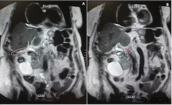
Spontaneous cholecystocutaneous fistula, one of the rarest complications of acute cholecystitis, has been reported in fewer than 25 cases over the past 50 years. Not only is this case rare but interestingly the patient experienced no pain or symptoms consistent with gallbladder pathology leading up to her hospitalisation. Furthermore, laboratory studies, microbiology and computed tomography scanning did not establish a diagnosis until the fistula passed calculi. An 85-year-old lady with multiple co-morbidities presented to the Emergency Department with an erythematous soft and non-tender mass in her right flank. The mass had spontaneously ruptured and was discharging a serous-like material. Prior to further investigation a working diagnosis of an eroding/fungating caecal tumour was made. The lesion continued to discharge over a 3 month period which heralded the passage of 11 small, brown calculi thought to be gallstones. At this point spontaneous cholecystocutaneous fistula was diagnosed and was later confirmed by magnetic resonance imaging cholangiopancreatography.
© JSCR.
Figures


References
-
- Vasanth A, Siddiqui A, O'Donnell K. Spontaneous cholecystocutaneous fistula. South Med J. 2004;97:183–5 - PubMed
-
- Nicholson T, Born MW, Garber E. Spontaneous cholecystocutaneous fistula presenting in the gluteal region. J Clin Gastroenterol. 1999;28:276–7 - PubMed
-
- Lyon C, Clarke DC. Diagnosis of acute abdominal pain in older patients. Am Fam Physician. 2006;74:1537–1544 - PubMed
LinkOut - more resources
Full Text Sources
Miscellaneous
