. 2014 Jun 20:10:25.
doi: 10.1186/1746-160X-10-25.

Dentin-like tissue formation and biomineralization by multicellular human pulp cell spheres in vitro

Affiliations

Dentin-like tissue formation and biomineralization by multicellular human pulp cell spheres in vitro

Jörg Neunzehn et al. Head Face Med. .

Abstract

Introduction: Maintaining or regenerating a vital pulp is a preferable goal in current endodontic research. In this study, human dental pulp cell aggregates (spheres) were applied onto bovine and human root canal models to evaluate their potential use as pre-differentiated tissue units for dental pulp tissue regeneration.

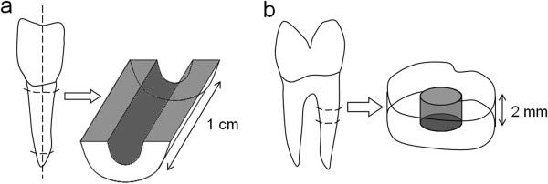
Methods: Human dental pulp cells (DPC) were derived from wisdom teeth, cultivated into three-dimensional cell spheres and seeded onto bovine and into human root canals. Sphere formation, tissue-like and mineralization properties as well as growth behavior of cells on dentin structure were evaluated by light microscopy (LM), confocal laser scanning microscopy (CLSM), scanning electron microscopy (SEM) and energy dispersive X-ray spectroscopy (EDX).

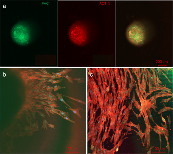
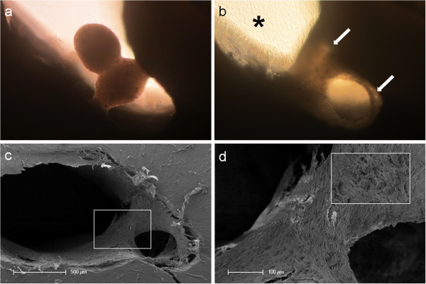
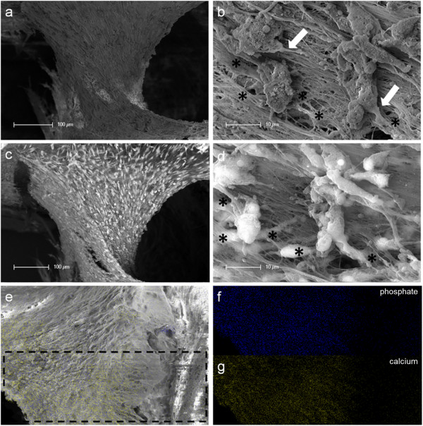
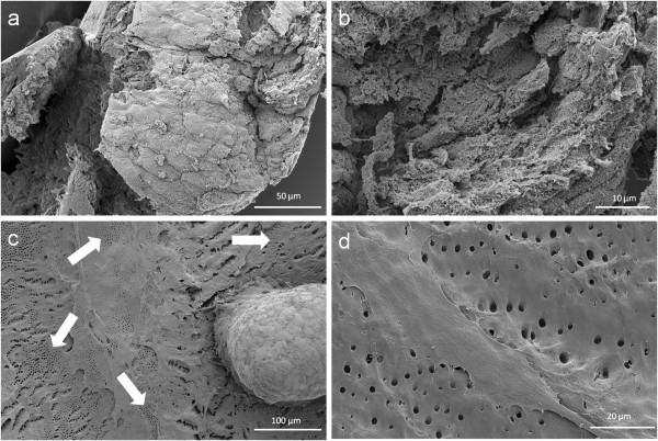
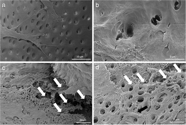
Results: Spheres and outgrown cells showed tissue-like properties, the ability to merge with other cell spheres and extra cellular matrix formation; CLSM investigation revealed a dense network of actin and focal adhesion contacts (FAC) inside the spheres and a pronounced actin structure of cells outgrown from the spheres. A dentin-structure-orientated migration of the cells was shown by SEM investigation. Besides the direct extension of the cells into dentinal tubules, the coverage of the tubular walls with cell matrix was detected. Moreover, an emulation of dentin-like structures with tubuli-like and biomineral formation was detected by SEM- and EDX-investigation.

Conclusions: The results of the present study show tissue-like behavior, the replication of tubular structures and the mineralization of human dental pulp spheres when colonized on root dentin. The application of cells in form of pulp spheres on root dentin reveals their beneficial potential for dental tissue regeneration.


Figures

Figure 1
Figure 1
Preparation of bovine and human root canal models. Bovine roots were sectioned longitudinally and the root canal surface was treated with a rose-bur (a). Additionally, human roots were cut horizontally into 2 mm thick discs after root canal preparation (b). Both models underwent ultrasonic desorption to remove the accumulated „smear layer“(abrasive dust, debris).
Figure 2
Figure 2
Confocal laser scanning microscopic evaluation. Actin (red), FAC (green) and DAPI (blue) stained cells in the upper third of a pulp sphere (a) and of its outgrown cells (b and c) representing a dense actin formation and consistent round cell nuclei.
Figure 3
Figure 3
Tissue-like behavior of human pulp sphere cells. Figure (a) shows merging pulp-spheres in a human root canal model after five days. After 28 days of cultivation the two spheres merged, cells grew out of the spheres and coated the root dentin walls (white arrows). (b) Outgrowing cells also covered the underlying polystyrene culture dish and built up a dense cell layer (black asterisk). SEM investigation of the merged spheres (c and d) represents the even surface of the new cell construct and the widespread coverage of the root canal walls (c) as well as the randomly distributed different sized particles (d).
Figure 4
Figure 4
SEM and EDX investigation of merged human pulp cell spheres in a human root canal. Figure (a) shows a fibrillar surface of the closed cell construct with different sized mineral like structures on it (Figure 4b) by detection of secondary electrons. The white arrows in figure (b) represent a cellular connection between the mineral particles and the underlying cell layer. The images in figure (c) and (d) show the same areas investigated by the detection of back scattered electrons with a clearly seen atomic number depending material contrast between the spheres’ cell matrix and the mineral particles. Black asterisks in figure (b) and (d) denote particles with higher atomic numbers, which could only be seen in figure (d) by the aid of the material contrast. The area of the sphere, which was investigated by EDX, is marked with black lines in figure (e). The results concerning the higher concentration of mineral specific elements are represented in figure (f) (phosphate) and figure (g) (calcium).
Figure 5
Figure 5
Scanning electron microscopic sphere and cell investigation. SEM images of the outer epithelial-like shell of a cell sphere (a) and the less organized cells with a higher production of extracellular matrix inside the sphere (b). Figure (c) shows a pulp sphere attached onto bovine dentin with a high number of outgrowing, multilayered cells as well as flat spread out and attached cells on bovine root dentin (d). The white arrows in figure (4c) highlight the dentin tubule-like holes in the cell multi-layer.
Figure 6
Figure 6
Cell behavior on bovine dentin. Scanning electron microscopic images show the bovine dentin surface orientated migration of outgrown pulp sphere cells (a) and the vortical ingrowth into the dentinal tubules as well as the covering of the tubular walls by these cells (b). Multilayered cell formations with cellular emulation of three-dimensional dentin structures could be detected in the multi layered outgrowth area adjacent to a sphere (white arrows in Figure 6c). The white arrows in figure (d) denote the fibrillar dentin tissue-like structure over numerous cell layers (black arrow).

References

    1. West J. Endodontic update 2006. J Esthet Restor Dent. 2006;18:280–300. - PubMed
    1. Zhang W, Yelick PC. Vital pulp therapy-current progress of dental pulp regeneration and revascularization. Int J Dent. 2010;2010:856087. - PMC - PubMed
    1. Randow K, Glantz PO. On cantilever loading of vital and non-vital teeth. An experimental clinical study. Acta Odontol Scand. 1986;44:271–277. - PubMed
    1. Tziafas D. The future role of a molecular approach to pulp-dentinal regeneration. Caries Res. 2004;38:314–320. - PubMed
    1. Murray PE, Garcia-Godoy F, Hargreaves KM. Regenerative endodontics: a review of current status and a call for action. J Endod. 2007;33:377–390. - PubMed

LinkOut - more resources