

Vulvar lipoma: a case report
- PMID: 24946809
- PMCID: PMC4077142
- DOI: 10.1186/1752-1947-8-203
Vulvar lipoma: a case report
Abstract
Introduction: Vulvar lipoma is a rare tumor localization and only a few cases have been reported. The clinical characteristics of vulvar lipoma are well known. However, it is important to distinguish lipomas from liposarcomas. We report a case of vulvar lipoma and discuss its clinical features, including diagnostic aspects, with emphasis on histopathological evaluation of all excised lesions. We also report and discuss patient management and treatment outcomes.
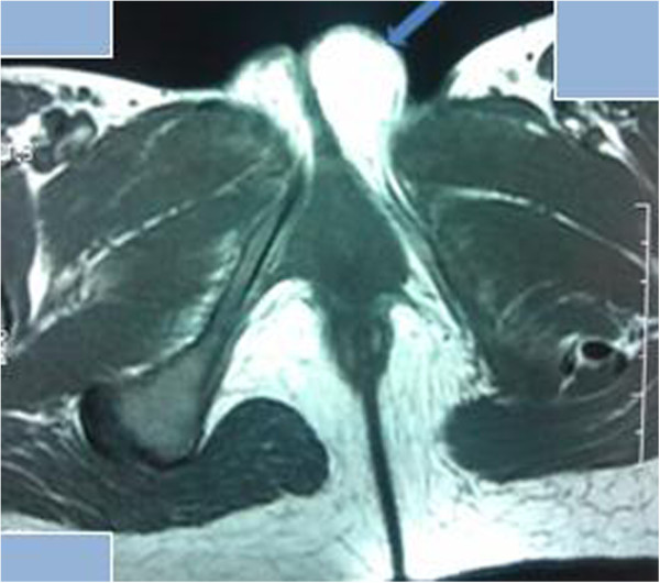
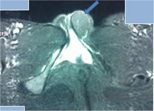
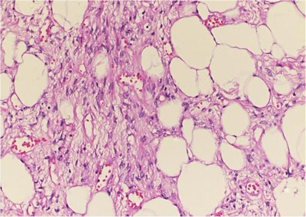
Case presentation: We report the case of a 27-year-old Moroccan woman. Our patient presented with a painless and slow-growing right vulvar mass that had evolved over one year, which had suddenly become uncomfortable when walking. A physical examination revealed a single soft and pasty mass in her left labium majus, which could be mobilized under her skin towards her mons pubis. The largest dimension of the mass measured 6cm. Magnetic resonance imaging showed a homogenous hyperintense mass with a well-defined contour in her left labium majus; a fat-suppressed magnetic resonance image demonstrated a marked signal intensity decrease. The mass was completely removed surgically. A histological examination revealed a circumscribed benign tumor composed of mature adipocytes, confirming the diagnosis of vulvar lipoma.
Conclusion: Vulvar lipomas must be differentiated from liposarcomas, which demonstrate very similar clinical and imaging profiles. The final diagnosis should be based on histopathological evaluation. A precise diagnosis should allow for appropriate surgical treatment.
Figures
References
-
- Jourjon R, Dohan A, Brouland JP, Guerrache Y, Fazel A, Soyer P. Angiolipoma of the labia majora: MR imaging findings with histopathological correlation. Clin Imaging. 2013;37:965–968. - PubMed
-
- Aust MC, Spies M, Kall S, Gohritz A, Boorboor P, Kolokythas P, Vogt PM. Lipomas after blunt soft tissue trauma: are they real? Analysis of 31 cases. Br J Dermatol. 2007;157(1):92–99. - PubMed
-
- Salam GA. Lipoma excision. Am Fam Physician. 2002;65(5):901–904. - PubMed
Publication types
MeSH terms
LinkOut - more resources
Full Text Sources
Other Literature Sources
Medical
