. 2014 Aug 15;74(16):4329-40.
doi: 10.1158/0008-5472.CAN-14-0093. Epub 2014 Jun 19.

IL4 receptor ILR4α regulates metastatic colonization by mammary tumors through multiple signaling pathways

Affiliations

IL4 receptor ILR4α regulates metastatic colonization by mammary tumors through multiple signaling pathways

Katherine T Venmar et al. Cancer Res. .

Abstract

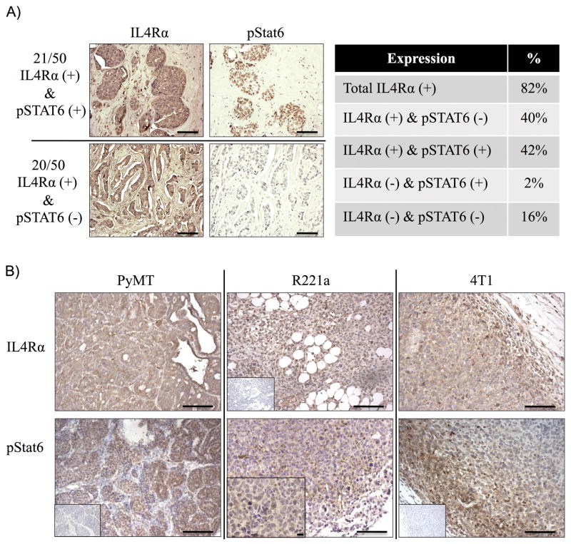
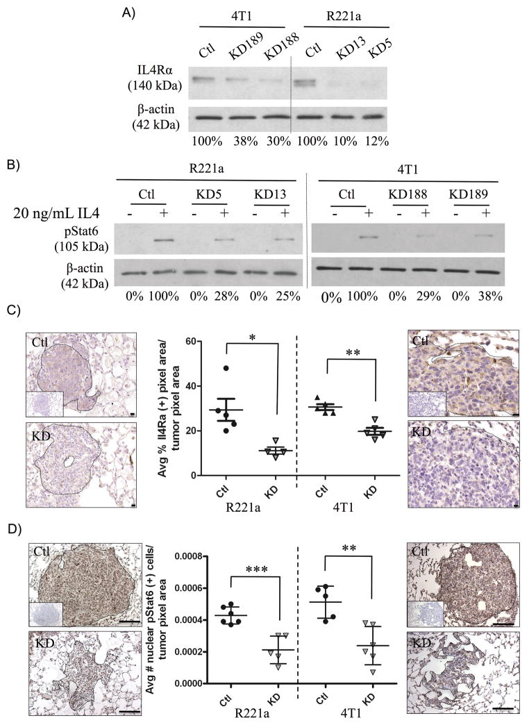
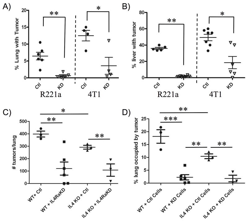
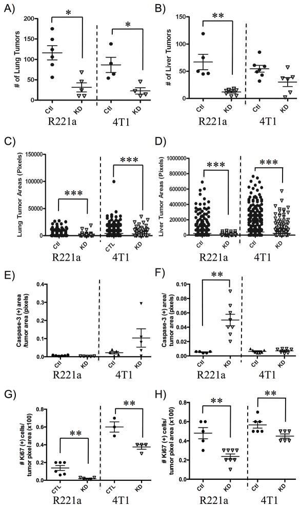
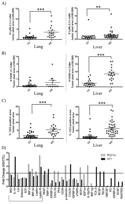
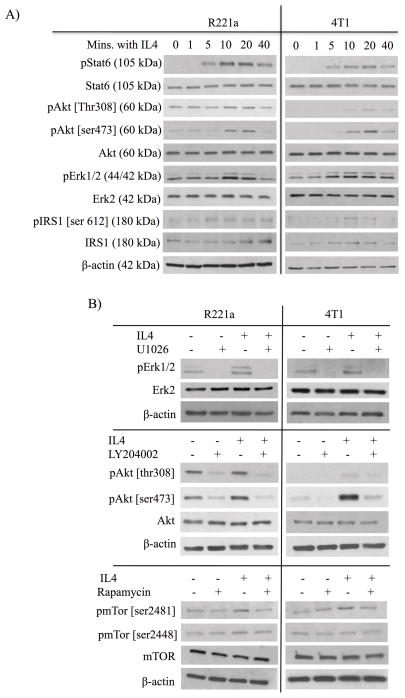
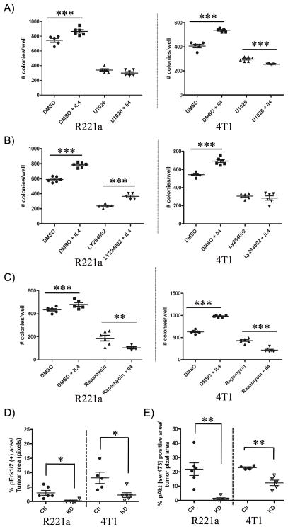
IL4, a cytokine produced mainly by immune cells, may promote the growth of epithelial tumors by mediating increased proliferation and survival. Here, we show that the type II IL4 receptor (IL4R) is expressed and activated in human breast cancer and mouse models of breast cancer. In metastatic mouse breast cancer cells, RNAi-mediated silencing of IL4Rα, a component of the IL4R, was sufficient to attenuate growth at metastatic sites. Similar results were obtained with control tumor cells in IL4-deficient mice. Decreased metastatic capacity of IL4Rα "knockdown" cells was attributed, in part, to reductions in proliferation and survival of breast cancer cells. In addition, we observed an overall increase in immune infiltrates within IL4Rα knockdown tumors, indicating that enhanced clearance of knockdown tumor cells could also contribute to the reduction in knockdown tumor size. Pharmacologic investigations suggested that IL4-induced cancer cell colonization was mediated, in part, by activation of Erk1/2, Akt, and mTOR. Reduced levels of pAkt and pErk1/2 in IL4Rα knockdown tumor metastases were associated with limited outgrowth, supporting roles for Akt and Erk activation in mediating the tumor-promoting effects of IL4Rα. Collectively, our results offer a preclinical proof-of-concept for targeting IL4/IL4Rα signaling as a therapeutic strategy to limit breast cancer metastasis.


Conflict of interest statement

No conflicts of interest to declare.

Figures

Figure 1
Figure 1. IL4 receptor expression and activation in human breast cancers and murine models
A) Left: Representative images of a human tissue microarray (TMA) of breast carcinoma samples (n = 50) stained by immunohistochemistry for IL4Rα or pSTAT6. Right: Summary of categorical (positive/negative) TMA staining results. B) Representative images of IL4Rα and pStat6 immunostaining in murine mammary tumors (n=3 each). Scale bar = 100 μM. Inset: negative staining controls, and one 40X image (scale bar = 10 μM) of pStat6 in an R221a tumor.
Figure 2
Figure 2. Functional knock down of IL4Rα expression in vitro and in vivo
A) Western blot illustrating the percent knockdown of IL4Rα protein expression and B) Stat6 activation in response to 20ng/mL IL4 in individual R221a and 4T1 control (ctl) and IL4Rα KD (KD) clones. C) Representative images and quantification of immunohistochemical staining for IL4Rα (40X, scale bar = 10 μM) or D) pStat6 (20X, scale bar = 100 μM) in R221a and 4T1 sh-control and IL4Rα KD lung metastases (n = 4–6). Inset: negative staining controls.
Figure 3
Figure 3. The IL4/IL4Rα interaction promotes metastatic tumor growth in vivo
Quantification of tumor burden from H&E stained A) lung and B) liver sections (3 per mouse) from mice receiving R221a or 4T1 IL4Rα KD or sh-control cells (lung: n = 4–6, liver: n = 5–8). C) Enumeration of lung surface tumors and D) quantification of lung tumor burden from H&E stained serial sections from Wild-type (WT) or IL4−/− (IL4 KO) mice receiving either 4T1 sh-control or IL4Rα KD clones by tail vein injection (n = 3–6).
Figure 4
Figure 4. IL4Rα-associated survival and proliferation contribute to metastatic tumor growth
A) Enumeration of R221a or 4T1 IL4Rα KD and sh-control lung and B) liver metastatic tumors per mouse (lung: n = 4–6, liver: n = 5–8) from H&E stained sections (3 per mouse). C) Quantification of individual lung or D) liver tumor areas from the same H&E sections. E) Quantification of total cleaved caspase-3 positive area per total IL4Rα KD or sh-control lung or F) liver tumor area for each mouse (lung: n = 4–6, liver: n = 5–8). G) Quantification of the total number of Ki67 positive cells per IL4Rα KD or sh-control tumor area for each mouse lung or H) liver (lung: n = 3–6, liver: n = 5–8).
Figure 5
Figure 5. IL4Rα expression is associated with changes in immune infiltration and cytokine production
Quantification of A) neutrophils, B) macrophages, and C) lymphocytes from immunostained lung and liver sections of mice receiving R221a or IL4Rα KD or sh-control cells (n = 4–7). D) Quantitation of the fold change in protein levels from R221a and 4T1 IL4Rα KD and sh-control conditioned media, assessed using a commercial cytokine array and densitometry. Proteins increased in IL4Rα KD clones for both cells lines that have potential to regulate immune recruitment are bracketed.
Figure 6
Figure 6. Inhibition of IL4-activated Stat6, Akt, and Erk
Representative western blots were completed using whole cell lysates from combined R221a or 4T1 sh-control clones, and 20 ng/mL IL4. A) Timecourse analysis of effector proteins activated within 40 minutes of IL4 treatment. B) U1026 (R221a: 10 μM; 4T1: 15 μM) inhibits IL4-activated pErk1/2, LY294002 (R221a: 5 μM; 4T1: 8 μM) inhibits IL4-activated pAkt, and rapamycin (R221a: 5 nM; 4T1 3 nM) inhibits pmTor.
Figure 7
Figure 7. The IL4/IL4Rα interaction promotes colonization ability via Erk, Akt, and mTor activation
R221a or 4T1 combined sh-control clones were seeded +/− IL4 (R221a: 2ng/mL, 4T1: 10 ng/mL) and +/− drug. A) U1026 (R221a: 10 μM, 4T1: 15 μM) inhibits IL4-induced colony formation. B) LY294002 inhibits colony formation in 4T1 cells (8 μM), but not R221a cells (5 μM). C) Rapamycin (R221a: 5 nM, 4T1 3 nM) inhibits IL4-induced colony formation. D) Quantification of pErk1/2 and E) pAkt [ser473] immunostaining in R221a and 4T1 IL4Rα KD and sh-control lung metastases. The percent positive area per mouse (n = 4–6) is shown.

References

    1. American Cancer Society. Cancer Facts & Figures 2013. Atlanta: American Cancer Society; 2013.
    1. Ben-Baruch A. Host microenvironment in breast cancer development: inflammatory cells, cytokines and chemokines in breast cancer progression: reciprocal tumor-microenvironment interactions. Breast Cancer Res. 2003;5:31–6. - PMC - PubMed
    1. Camp BJ, Dyhrman ST, Memoli VA, Mott LA, Barth RJ. In situ cytokine production by breast cancer tumor-infiltrating lymphocytes. Ann Surg Oncol. 1996;3:76–84. - PubMed
    1. Todaro M, Lombardo Y, Francipane MG, Alea MP, Cammareri P, Iovino F, et al. Apoptosis resistance in epithelial tumors is mediated by tumor-cell-derived interleukin-4. Cell Death Differ. 2008;15:762–72. - PubMed
    1. Nelms K, Keegan AD, Zamorano J, Ryan JJ, Paul WE. The IL-4 receptor: signaling mechanisms and biologic functions. Ann Rev Immunol. 1999;17:701–38. - PubMed

Publication types

Substances