. 2014 Aug 1;289(31):21640-50.
doi: 10.1074/jbc.M114.578765. Epub 2014 Jun 19.

Evidence supporting the 19 β-strand model for Tom40 from cysteine scanning and protease site accessibility studies

Affiliations

Evidence supporting the 19 β-strand model for Tom40 from cysteine scanning and protease site accessibility studies

Sebastian W K Lackey et al. J Biol Chem. .

Abstract

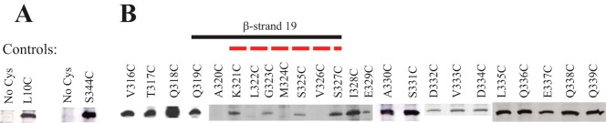
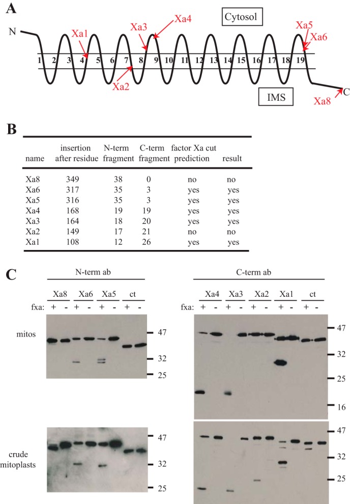
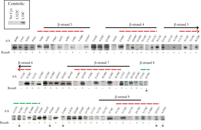
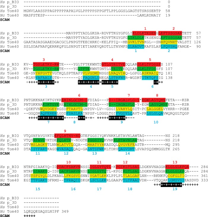
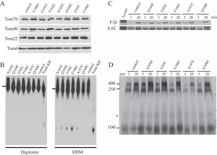
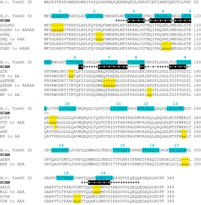
Most proteins found in mitochondria are translated in the cytosol and enter the organelle via the TOM complex (translocase of the outer mitochondrial membrane). Tom40 is the pore forming component of the complex. Although the three-dimensional structure of Tom40 has not been determined, the structure of porin, a related protein, has been shown to be a β-barrel containing 19 membrane spanning β-strands and an N-terminal α-helical region. The evolutionary relationship between the two proteins has allowed modeling of Tom40 into a similar structure by several laboratories. However, it has been suggested that the 19-strand porin structure does not represent the native form of the protein. If true, modeling of Tom40 based on the porin structure would also be invalid. We have used substituted cysteine accessibility mapping to identify several potential β-strands in the Tom40 protein in isolated mitochondria. These data, together with protease accessibility studies, support the 19 β-strand model for Tom40 with the C-terminal end of the protein localized to the intermembrane space.

Keywords: Membrane Protein; Mitochondria; Neurospora; Protein Structure; SCAM; Tom40; Transmembrane Domain; β-Barrel Protein.


Figures

FIGURE 1.
FIGURE 1.
Defining the position of the C-terminal β-strand of NcTom40. A, SCAM analysis of control strains. Strains were developed that expressed Tom40 protein that lacked any Cys residues (No Cys) or contained single Cys residues near the N or C terminus of the protein (L10C and S344C) at positions predicted to be labeled by the maleimide reagent. Mitochondria were isolated from the strains and exposed to the labeling reagent, Tom40 was immunoprecipitated, and Western blot analysis using streptavidin-HRP to detect biotin maleimide-labeled Tom40 molecules was performed as described under “Experimental Procedures.” The appearance of a band at a given position indicates that the Cys residue is present in an aqueous environment and is able to react with the biotin maleimide. B, as in A, except that mitochondria for SCAM analysis were isolated from strains expressing Tom40 with single Cys substitution mutations at the indicated positions. The region that conforms to the predicted alternating labeling pattern for β-strand 19 is indicated by a solid black line. The region predicted as β-strand 19 for NcTom40 by modeling (12) is shown by a red dotted line. The results for the Cys mutant strains were not all derived from the same gel/Western blot. Controls were present on each gel/blot, but these and other irrelevant lanes have been removed to allow linear presentation of data.
FIGURE 2.
FIGURE 2.
Protease accessibility of triple factor Xa sites introduced into NcTom40. A, representation of NcTom40 in the outer membrane with the relative position of predicted cytosolic loops, membrane spanning β-strands (numbered), and IMS loops shown as predicted for the 19-strand model (12). The locations of triple factor Xa sites introduced into the protein in individual strains are indicated by red arrows. B, the sites where triple factor Xa sites were inserted into the NcTom40 protein, the predicted sizes (kDa) of the N- and C-terminal (N-term and C-term) fragments that would be produced by cleavage at those sites, and the prediction of cleavage by factor Xa treatment of intact mitochondria, based on the 19 β-strand structural model, are shown. The predictions assume that the C terminus exists in the IMS and that triple factor Xa sites beginning within a β-strand have at least one or two of the Xa sites exposed in the compartment that is at the C-terminal end of the insertion site. The actual result of cleavage by factor Xa protease, as shown in panel C, is also given. C, the two top panels show Western blots of isolated, mitochondria (mitos) containing Tom40 protein with factor Xa sites inserted at the positions indicated in panel A. Mitochondria were treated with factor Xa protease (fxa) or left untreated (as indicated) and then subjected to SDS-PAGE and Western blotting. The two lower panels are identical to the upper panels, except that the mitochondria were subjected to a procedure that creates crude mitoplasts, prior to treatment with factor Xa. This allows the protease access to the IMS and exposes both sides of the outer membrane to the protease. The two left panels were immunodecorated with antibody produced to a peptide corresponding to the first 12 amino acids of NcTom40 (N-term ab). The two right panels were decorated with antibody produced to a peptide for the last 12 amino acids of NcTom40 (C-term ab). The ct lanes are mitochondria isolated from a control strain (76-26) containing no factor Xa sites. The positions of standard molecular weight markers are shown on the right side of the blots. The C-terminal antibody detects an unknown band in certain lanes, just below the full-length proteins.
FIGURE 3.
FIGURE 3.
SCAM analysis of different regions of NcTom40. Results and presentation are as described in the legend to Fig. 1. Controls are shown in the upper left box and include L322C as a negative control based on the results in Fig. 1B. β-Strands are as indicated as in Fig. 1. The arrowheads for β-strands 5 and 6 indicate that the strand should extend further in the indicated direction, but no SCAM data were obtained for the region. The labeling result is shown below each position. * indicates positions where labeling was expected, due to the position of a predicted strand or loop, but not observed. The dashed green line indicates the predicted position of β-strand 8 (12) that was not confirmed by experimental results (see text).
FIGURE 4.
FIGURE 4.
Alignment of human porin, human Tom40, and NcTom40 with SCAM results. The amino acid sequence of porin from Homo sapiens (Hs) is shown in the top two sequences. In the first (Hs p_BIO) the position of the 13 β-strands predicted by the BIO model (36) are shown (highlighted and numbered in red). An asterisk (*) represents a sharp (zero residue) hairpin loop between β-strands 7 and 8 in the BIO model. The second sequence (Hs p_3D) shows the position (green) of the 19 β-strands from the three-dimensional structures of porin. The third sequence (Hs Tom40) shows the position (yellow) of the 19 β-stands predicted to exist in human Tom40 (11–13). The bottom sequence (Nc Tom40) represents the NcTom40 showing the predicted 19 β-strands (highlighted and numbered in teal). The position and alignment of strands in the last three sequences are as previously described (12). The SCAM results from this study are displayed below the NcTom40 sequence. Positive (+) and negative (−) labeling signals are indicated. SCAM results that suggest the position of a β-strand are shown as highlighted black bars below the sequence.
FIGURE 5.
FIGURE 5.
Analysis of TOM complex composition and function in mitochondria from selected Tom40 Cys-mutant strains. A, cells from the indicated strains (top of panel) were grown in liquid culture, and mitochondria were isolated. Mitochondrial proteins were subjected to SDS-PAGE followed by transfer to nitrocellulose, and immunodecoration with the antibodies indicated on the left. The control strain was 76-26, which contains wild type Tom40. B, isolated mitochondria from the strains indicated (top of panel) were dissolved in 1% digitonin or 1% n-dodecyl-β-d-maltoside (DDM) for analysis of complex stability using BNGE. Blots were decorated with Tom40 antibody. The controls are Tom6 RIP (a strain lacking Tom6 that shows severely reduced TOM complex stability (51)) and 76-26 (control), which contains wild type TOM complex. C, radiolabeled matrix precursor F1β and inner membrane precursor ADP/ATP carrier (AAC) were incubated (for 5 or 20 min, as indicated) with mitochondria isolated from the Tom40 Cys-substitution strains or the 76-26 control. Following import, mitochondria were subjected to SDS-PAGE. Proteins were transferred to nitrocellulose membrane, and import was analyzed by autoradiography. Lysate represents 33% of the radiolabeled lysate added to each import reaction. D, radiolabeled Tom40 precursor was incubated for 5 and 20 min with mitochondria isolated from the Tom40 Cys-substitution strains indicated (top of panel) as well as the 76-26 control strain. Mitochondria were dissolved in 1% digitonin and subjected to BNGE. The proteins were transferred to PVDF membrane and analyzed by autoradiography. The size of the mature TOM complex (400 kDa), and assembly intermediates I (250 kDa) and II (100 kDa) are indicated on the left. * indicates an undefined band.
FIGURE 6.
FIGURE 6.
Previously studied mutants of N. crassa Tom40 aligned with the three-dimensional model and SCAM results. The top sequence is the wild type NcTom40 protein with the 19 modeled β-strands shown (highlighted and numbered in teal). All SCAM results (from Figs. 1 and 3) are indicated below the NcTom40 sequence. Black shading indicates the regions with alternating label (+) and no label (−) signals that are likely to represent β-strands. Below the SCAM results is a depiction of the Tom40 protein of each mutant with the region that is discussed in the text containing the amino acid deletions (−) or alanine substitutions (a) indicated (19, 40). The region affected in each mutant is highlighted in yellow.

Similar articles

Cited by

References

    1. Neupert W., Herrmann J. (2007) Translocation of proteins into mitochondria. Annu. Rev. Biochem. 76, 723–749 - PubMed
    1. Endo T., Yamano K. (2009) Multiple pathways for mitochondrial protein traffic. Biol. Chem. 390, 723–730 - PubMed
    1. Chacinska A., Koehler C. M., Milenkovic D., Lithgow T., Pfanner N. (2009) Importing mitochondrial proteins: machineries and mechanisms. Cell 138, 628–644 - PMC - PubMed
    1. Schmidt O., Pfanner N., Meisinger C. (2010) Mitochondrial protein import: from proteomics to functional mechanisms. Nat. Rev. Mol. Cell Biol. 11, 655–667 - PubMed
    1. Becker T., Böttinger L., Pfanner N. (2012) Mitochondrial protein import: from transport pathways to an integrated network. Trends Biochem. Sci. 37, 85–91 - PubMed

Publication types

MeSH terms

LinkOut - more resources