. 2014 Aug 15;289(33):22926-22941.
doi: 10.1074/jbc.M114.579672. Epub 2014 Jun 19.

Actin enables the antimicrobial action of LL-37 peptide in the presence of microbial proteases

Affiliations

Actin enables the antimicrobial action of LL-37 peptide in the presence of microbial proteases

Asaf Sol et al. J Biol Chem. .

Abstract

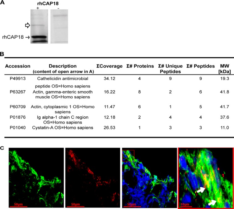
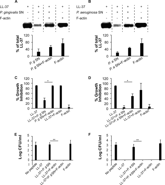
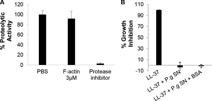
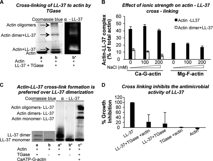
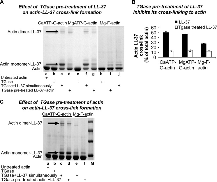
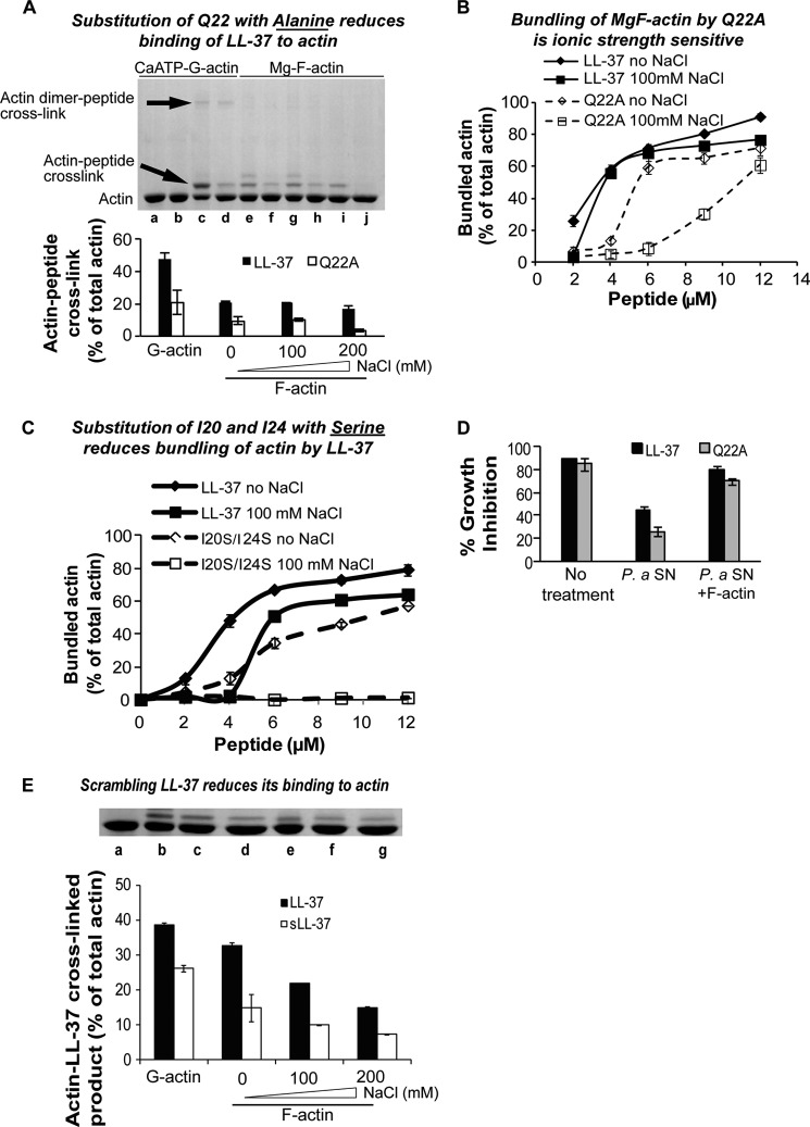
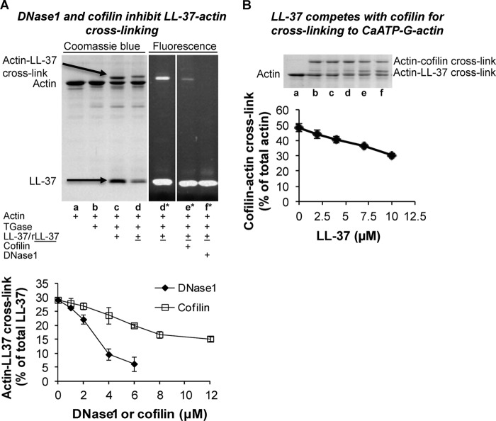
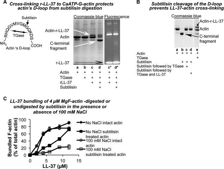
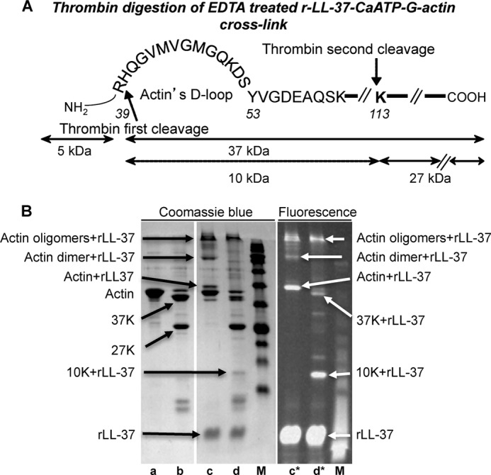
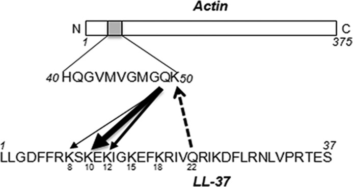
Host defense peptides play an important host-protective role by their microcidal action, immunomodulatory functions, and tissue repair activities. Proteolysis is a common strategy of pathogens used to neutralize host defense peptides. Here, we show that actin, the most abundant structural protein in eukaryotes, binds the LL-37 host defense peptide, protects it from degradation by the proteases of Pseudomonas aeruginosa and Porphyromonas gingivalis, and enables its antimicrobial activity despite the presence of the proteases. Co-localization of LL-37 with extracellular actin was observed in necrotized regions of samples from oral lesions. Competition assays, cross-linking experiments, limited proteolysis, and mass spectrometry revealed that LL-37 binds by specific hydrophobic interactions to the His-40-Lys-50 segment of actin, located in the DNase I binding loop. The integrity of the binding site of both LL-37 and actin is a prerequisite to the binding. Our results demonstrate that actin, presumably released by dead cells and abundant in infected sites, might be utilized by the immune system to enhance spatio-temporal immunity in an attempt to arrest infection and control inflammation.

Keywords: Actin; Antimicrobial Peptide (AMP); Host Defense; Protease; Transglutaminase.


Figures

FIGURE 1.
FIGURE 1.
Actin binds rhCAP18 and LL-37. Coomassie Blue-stained SDS-PAGE (A) and mass spectrometry analysis (B) of salivary factors (open arrow) that were pulled down (see “Experimental Procedures”) with Ni-NTA-agarose beads coated (+) or not (−) with rhCAP18 (closed arrow) used as bait. C, co-localization of LL-37 (red) with extracellular actin (green). Confocal microscopy imaging of immunofluorescence (see under “Experimental Procedures”) was performed on a sample of granular tissue collected from a human oral lesion. The merged channel contains nuclei staining with Hoechst (blue). Right panel shows the inset indicated in the merged image. Arrows point to co-localization of actin and LL-37.
FIGURE 2.
FIGURE 2.
F-actin protects and enables the antibacterial activity of LL-37 in the presence of bacterial proteases. A and B, Western immunodetection and densitometry analysis (mean ± S.D. of two independent experiments) of 3 μm LL-37 incubated with 5 μl of culture supernatant (SN) of P. gingivalis (P.g) (A) or P. aeruginosa (P.a) (B), in the absence or presence of 9 μm F-actin. C and D, growth inhibition of B. subtilis by 1.1 μm LL-37 treated or not with P. gingivalis (P.g) (C) or P. aeruginosa (P.a) (D) supernatant (SN), in the presence or absence of 3 μm F-actin. E and F, killing of B. subtilis (displayed as colony-forming units, CFU) by 1.1 μm LL-37 treated or not with P. gingivalis (P.g) (E) or P. aeruginosa (P.a) (F) supernatant (SN) in the presence or absence of 3 μm F-actin. * represents p < 0.05 in Student's t test, ** represents p < 0.01 in one-way analysis of variance.
FIGURE 3.
FIGURE 3.
Prevention of LL-37 proteolysis by microbial proteases is mediated by protection rather than protease inhibition. A, actin does not inhibit the proteolytic activity of R-gingipain of P. gingivalis (P.g). Protease activity (quantified using BAPNA as an R-gingipain substrate, see “Experimental Procedures”) of P. gingivalis conditioned medium co-incubated with or without 3 μm F-actin or with 1 μl of protease inhibitor mixture used as control. B, unlike actin, BSA (see Fig. 2C) does not protect the antimicrobial activity of LL-37 from the proteases of P. gingivalis. Presented data are mean ± S.D. of two independent experiments performed in triplicate. SN, supernatant.
FIGURE 4.
FIGURE 4.
Binding of LL-37 to the bacterial membrane is maintained in the presence of F-actin. FACS histogram (representative experiment) (A) and analysis of three independent experiments (B) of B. subtilis PY79 labeling when incubated with 1.1 μm tetramethylrhodamine-labeled LL-37, in the presence (blue dashed in A) or absence (red in A) of 3 μm F-actin, are shown. Gray histogram in A represents the background of B. subtilis without LL-37.
FIGURE 5.
FIGURE 5.
Cross-linking of LL-37 to actin by TGase. A, SDS-PAGE of cross-linking of 10 μm LL-37 to 8 μm CaATP-G-actin by 0.2 mg/ml TGase. Lane a, actin only; lane b, actin cross-linked to LL-37 (see “Experimental Procedures”) visualized by Coomassie Blue (left) and by Western immunodetection (right, lane b*). B, effect of NaCl concentration on 0.2 mg/ml TGase-induced cross-link formation between 4 μm CaATP-G or Mg-F-actin and 9 μm LL-37 (mean ± S.D. of three independent experiments). C, actin-LL-37 cross-link formation is preferred over LL-37 dimerization. 66 μm LL-37 were incubated with (lanes a and c) and without (lane b) 0.4 mg/ml TGase in the absence (lane b) and in the presence (lane c) of 60 μm CaATP-G-actin at room temperature for 2 h (lanes a and b), Coomassie blue stain; lane a*, lane b*, and lane c*, Western immunodetection. D, antimicrobial activity of LL-37 is lost upon formation of LL-37-LL-37 or LL-37-actin covalent cross-links. 1.1 μm LL-37 was incubated with or without 1.1 μm F-actin and with 0.4 mg/ml TGase at room temperature for 2 h. Following incubation samples were added to B. subtilis cells in a 96-well plate, and antimicrobial activity was measured as described under “Experimental Procedures.”
FIGURE 6.
FIGURE 6.
Pretreatment of LL-37 (but not of actin) with TGase inhibits actin-LL-37 cross-linking. A, 8 μm LL-37 was reacted with 0.2 mg/ml TGase at room temperature for 2 h and then cross-linked with 4 μm CaATP-G-actin, MgATP-G-actin, or Mg-F-actin for an additional 2 h. Lane a, actin alone; lanes b, e, and h, actin and TGase; lanes c, f, and j, LL-37 and TGase added simultaneously to actin and incubated for 2 h; lanes d, g, and i, LL-37 was preincubated for 2 h at room temperature with TGase before adding it to actin. B, effect of LL-37 pretreatment with TGase on the LL-37-actin cross-link formation. 82 μm LL-37 were treated with 1.8 mg/ml TGase at room temperature for 2 h and then 9 μm treated or untreated LL-37 and 0.2 mg/ml TGase was added to 4 μm actin and incubated for an additional 2 h (mean ± S.D. of three independent experiments). C, effect of actin pretreatment with TGase on the LL-37-actin cross-link formation. 8 μm actin was incubated with 0.2 mg/ml TGase on ice for 24 h and then 10 μm LL-37 was added, and the incubation was continued for an additional 24 h. Lanes a–d, CaATP-G-actin; lanes e and f, MgF-actin. Lane a, untreated actin; lane b, TGase-pretreated actin, no LL-37; lanes c and e, TGase pretreated actin with LL-37; lanes d and f, untreated actin with LL-37 and TGase added simultaneously; M, molecular weight marker.
FIGURE 7.
FIGURE 7.
Replacing LL-37's Gln-22 with alanine and Ile-20 and Ile-24 with serine, and scrambling reduces binding of LL-37 to actin. A, cross-linking of 4 μm actin with 9 μm Q22A or LL-37 at low ionic strength and in the presence of 100 and 200 mm NaCl. Upper panel, SDS-PAGE. Lower panel, densitometry analysis. Lanes a–d, CaATP-G-actin; lanes e–j, MgF-actin. Lane a, G-actin alone; lane b, G-actin and TGase, no LL-37; lane c, G-actin, LL-37, and TGase; lane d, G-actin, Q22A, and TGase; lane e, F-actin, LL-37, and TGase; lane f, F-actin, Q22A, and TGase; lane g, F-actin, LL-37, TGase, and 100 mm NaCl; lane h, F-actin, Q22A, TGase, and 100 mm NaCl; lane i, F-actin, LL-37, TGase, and 200 mm NaCl; lane j, F-actin, Q22A, TGase, and 200 mm NaCl. B, bundling of 4 μm MgF-actin by 2–12 μm LL-37 and Q22A at low ionic strength and in the presence of 100 mm NaCl. C, bundling of 4 μm MgF-actin by 3–20 μm I20S/I24S and LL-37 at low ionic strength and in the presence of 100 mm NaCl. For bundling procedure see “Experimental Procedures.” D, F-actin protects the antimicrobial activity of Q22A from proteolysis. Antimicrobial activity of LL-37 and of Q22A and their protection assay was performed as described under “Experimental Procedures.” SN, supernatant. E, cross-linking of 4 μm CaATP-G-actin and MgF-actin with 9 μm LL-37 or s-LL37 in the presence of increasing concentrations of NaCl. s-LL-37 or LL-37 was incubated simultaneously with 4 μm actin and 0.2 mg/ml TGase. Upper panel, SDS-PAGE, Lane a, actin only; lane b, actin, LL-37, TGase; lane c, actin, LL-37, TGase, 100 mm NaCl; lane d, actin, LL-37, TGase, 200 mm NaCl; actin, s-LL-37, TGase; lane e, actin, s-LL-37, TGase; lane f, actin, s-LL-37, TGase, 100 mm NaCl; lane g, actin, s-LL-37, TGase, 200 mm NaCl. Lower panel, quantitative assessment of LL-37-actin cross-link obtained from densitometry of SDS-PAGE.
FIGURE 8.
FIGURE 8.
DNase I and cofilin inhibit cross-linking of LL-37 to actin. A, representative gel (upper panel) and quantitative assessment (lower panel) of CaATP-G-actin (4 μm) cross-linked (“Experimental Procedures”) with 9 μm r-LL-37 or LL-37 by 0.2 mg/ml TGase in the presence and absence of 6 μm DNase I or 12 μm cofilin. All the constituents were incubated simultaneously at room temperature for 90 min. Lane a, actin alone; lane b, actin, TGase; lane c, actin, LL-37, TGase; lane d, actin, r-LL-37, TGase; lane e, actin, r-LL-37, cofilin, TGase; lane f, actin, r-LL-37, DNase I, TGase; *, rhodamine fluorescence. Samples were treated as in Fig. 6B. Evaluation is based on densitometry of SDS-PAGE. B, competition between 8 μm cofilin and 0–10 μm LL-37 for cross-linking to 4 μm CaATP-G-actin by TGase. LL-37 and cofilin were added simultaneously to CaATP-G-actin. For cross-linking procedure, see “Experimental Procedures.” Upper panel, SDS-PAGE. Lane a, actin only; lane b, actin, cofilin, TGase; lane c, actin, cofilin, TGase, 2 μm LL-37; lane d, actin, cofilin, TGase, 4 μm LL-37; lane e, actin, cofilin, TGase, 7 μm LL-37; lane f, actin, cofilin, TGase, 10 μm LL-37. Lower panel, quantitative evaluation of cross-linking of cofilin with actin from densitometry of SDS-PAGE.
FIGURE 9.
FIGURE 9.
Analysis of actin's LL-37-binding site by subtilisin digestion. A, cross-linking 6 μm r-LL-37 to 4 μm CaATP-G-actin protects actin's D-loop from subtilisin digestion. TGase reaction was stopped by 1 mm cysteamine; 30 min digestion with 1 μg/ml subtilisin was stopped by 1 mm PMSF; incubations were performed at room temperature. Samples were separated on SDS-PAGE and visualized by Coomassie Blue staining (lanes a–d) or by tetramethylrhodamine fluorescence (lanes c* and d*). Lane a, actin alone; lane b, actin and TGase; lanes c and c*, actin, r-LL-37, and TGase (added simultaneously); lanes d and d*, actin, r-LL-37, and TGase added simultaneously and followed by subtilisin digestion. Illustration on the left shows cleavage of CaATP-G-actin between Met-47 and Gly-48 by subtilisin (small black arrow) and covalent binding between Gln-41 and Lys-50 by TGase (two- headed arrow). B, subtilisin cleavage of the D-loop prevents LL-37-actin cross-linking. Other than using 9 μm LL-37, the same conditions as in A were applied. Lane a, actin; lane b, subtilisin-digested actin; lane c, subtilisin-digested actin treated by TGase; lane d, subtilisin-digested actin treated by TGase and then incubated with LL-37 for an additional 90 min; lane e, actin incubated with LL-37 and TGase (added simultaneously) for 90 min. C, LL-37 bundling of 4 μm MgF-actin digested or undigested by subtilisin in the presence or absence of 100 mm NaCl. 50 μm CaATP-G-actin were digested by subtilisin (at a 168:1 w/w/ratio) for 30 min, stopped by 1 mm PMSF and polymerized to MgF-actin by 2 mm MgCl2 (subtilisin digested F-actin). To 4 μm subtilisin, digested F-actin LL-37 was added in increasing concentrations, and bundling was carried out as described under “Experimental Procedures.”
FIGURE 10.
FIGURE 10.
LL-37 binds the actin His-40–Lys-113 10-kDa segment. A, illustration of thrombin digestion of EDTA-treated CaATP-G-actin. B, thrombin digestion of EDTA-treated r-LL-37-CaATP-G-actin cross-link. Lane a, 8 μm CaATP-G-actin was incubated with 1.5 mm EDTA for 21 h; lane b, same as lane a, but 30 μg/ml thrombin was also added together with EDTA, and proteolysis was stopped by 1 mm PMSF; lane c, 8 μm CaATP-G-actin cross-linked with 16 μm r-LL-37 by 0.3 mg of TGase for 2 h; the reaction was stopped by 1 mm cysteamine, and 1.5 mm EDTA was added followed by incubation for 21 h; lane d, same as lane c, but 30 μg/ml thrombin was also added together with EDTA, and proteolysis was stopped as in lane b; M, molecular weight marker. All incubations were performed at room temperature. Samples were separated on SDS-PAGE and visualized by Coomassie Blue stain (left panel) or by fluorescence (right panel, lanes c* and d*).
FIGURE 11.
FIGURE 11.
Identification of the residues involved in cross-linking between actin and LL-37 by mass spectrometry. A, model of the F-actin molecule (green) and the D-loop (gray sticks) using the PyMOL software. Enlarged illustration shows the location of Gln-49 in the D-loop. B and C, relative abundance of the D-loop sequences in actin and in LL-37-actin cross-linked bands. B, His-40–Lys-50 peptide is practically missing from the digest of the actin-LL-37 cross-linked heterodimer, which indicates its involvement in the cross-link formation. C, quantity of VMVGMGQK peptide in the His-40–Lys-50 sequence, containing Gln-49, is greatly reduced in the digest of actin-LL-37 cross-linked heterodimer relative to its quantity in the actin digest. D and E, mass spectrometry analysis and relative abundance of selected peptides in the digest of LL-37-actin or Q22A-actin cross-linked heterodimers. D, mass spectrometry relative abundance analysis show that Gln-22 and five lysines (Lys-8, Lys-10, Lys-12, and to a lesser extent Lys-15 and Lys-18) in LL-37 can participate in the formation of LL-37-actin cross-link. E, similar analysis of the Q22A-actin cross-linked digest indicates the participation of the same lysine residues in the cross-linking reaction. The residues marked in red could participate in the cross-link reaction. A change in font size indicates the significance of the residues in the cross-linking according to the MS calculation of peak area ratio between LL-37 or Q22A and actin-LL-37 or Q22A cross-link.
FIGURE 12.
FIGURE 12.
Schematic model of the significant residues that participate in cross-linking of LL-37 and actin by TGase. Arrow thickness signifies the residue's involvement in the cross-linking.

Similar articles

Cited by

References

    1. Altman H., Steinberg D., Porat Y., Mor A., Fridman D., Friedman M., Bachrach G. (2006) In vitro assessment of antimicrobial peptides as potential agents against several oral bacteria. J. Antimicrob. Chemother. 58, 198–201 - PubMed
    1. Hilpert K., Volkmer-Engert R., Walter T., Hancock R. E. (2005) High-throughput generation of small antibacterial peptides with improved activity. Nat. Biotechnol. 23, 1008–1012 - PubMed
    1. Radzishevsky I. S., Rotem S., Bourdetsky D., Navon-Venezia S., Carmeli Y., Mor A. (2007) Improved antimicrobial peptides based on acyl-lysine oligomers. Nat. Biotechnol. 25, 657–659 - PubMed
    1. Hancock R. E., Nijnik A., Philpott D. J. (2012) Modulating immunity as a therapy for bacterial infections. Nat. Rev. Microbiol. 10, 243–254 - PubMed
    1. Brogden K. A. (2005) Antimicrobial peptides: pore formers or metabolic inhibitors in bacteria? Nat. Rev. Microbiol. 3, 238–250 - PubMed

Publication types

MeSH terms

LinkOut - more resources