. 2014 Aug;34(8):1723-30.
doi: 10.1161/ATVBAHA.114.303983. Epub 2014 Jun 19.

Transplanted perivascular adipose tissue accelerates injury-induced neointimal hyperplasia: role of monocyte chemoattractant protein-1

Affiliations

Transplanted perivascular adipose tissue accelerates injury-induced neointimal hyperplasia: role of monocyte chemoattractant protein-1

David Manka et al. Arterioscler Thromb Vasc Biol. 2014 Aug.

Abstract

Objective: Perivascular adipose tissue (PVAT) expands during obesity, is highly inflamed, and correlates with coronary plaque burden and increased cardiovascular risk. We tested the hypothesis that PVAT contributes to the vascular response to wire injury and investigated the underlying mechanisms.

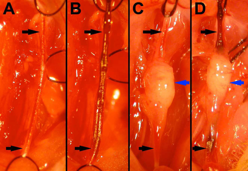
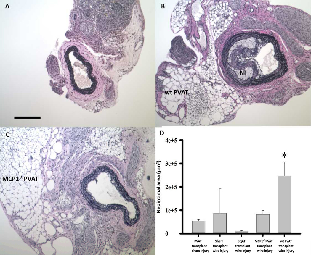
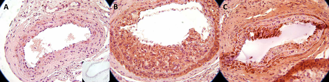
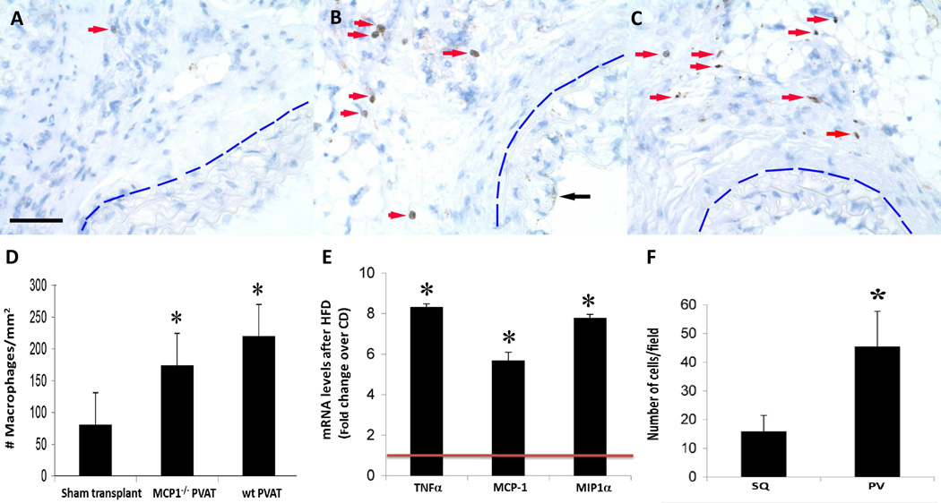
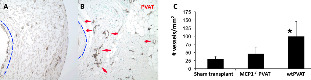
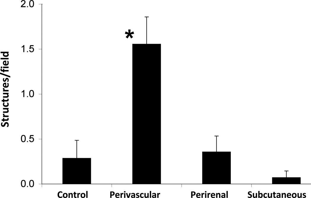
Approach and results: We transplanted thoracic aortic PVAT from donor mice fed a high-fat diet to the carotid arteries of recipient high-fat diet-fed low-density lipoprotein receptor knockout mice. Two weeks after transplantation, wire injury was performed, and animals were euthanized 2 weeks later. Immunohistochemistry was performed to quantify adventitial macrophage infiltration and neovascularization and neointimal lesion composition and size. Transplanted PVAT accelerated neointimal hyperplasia, adventitial macrophage infiltration, and adventitial angiogenesis. The majority of neointimal cells in PVAT-transplanted animals expressed α-smooth muscle actin, consistent with smooth muscle phenotype. Deletion of monocyte chemoattractant protein-1 in PVAT substantially attenuated the effects of fat transplantation on neointimal hyperplasia and adventitial angiogenesis, but not adventitial macrophage infiltration. Conditioned medium from perivascular adipocytes induced potent monocyte chemotaxis in vitro and angiogenic responses in cultured endothelial cells.

Conclusions: These findings indicate that PVAT contributes to the vascular response to wire injury, in part through monocyte chemoattractant protein-1-dependent mechanisms.

Keywords: adipose tissue; hyperplasia.


Figures

Figure 1
Figure 1
Surgical images taken during wire injury procedure. Two weeks after either sham transplantation (A, B) or transplantation of 2–3mg of perivascular adipose tissue (C, D), wire injury was performed (B,D). The carotid artery (black arrows) was ligated with silk sutures proximally and distally, relative to the carotid bifurcation. Note that transplanted PVAT is healthy appearing, with incorporated vessels, at the time of wire injury (blue arrows, C & D).
Figure 2
Figure 2
Carotid arteries from LDLR−/− mice following wire injury. Compared to sham-transplanted control (A), transplanted wild-type (wt) PVAT resulted in a robust neointimal response (NI, panel B). Note that the transplanted adipocytes are healthy appearing and in intimate contact with the lamina adventitia. Transplanted MCP-1−/− PVAT was not associated with robust NI formation (C). scale bar = 200µm. (D) Transplanted PVAT increases cross-sectional area of neointimal lesion following wire injury. *p<0.05 PVAT + injury vs. all other groups. Data were analyzed by one-way analysis of variance (ANOVA) followed by pairwise multiple comparison procedures (Holm-Sidak method).
Figure 3
Figure 3
Composition of VSMC in neointima following PVAT transplantation and wire injury. SM actin staining in sham-transplanted control (A), transplanted wild-type (wt) PVAT (B) and transplanted MCP-1−/− PVAT (C) carotid arteries following wire injury. Note that the majority of NI cells in B and C stain positively for SMA.
Figure 4
Figure 4
Inflammation following PVAT transplantation and wire injury. F4/80 staining revealed few adventitial macrophages following wire injury surrounding sham-transplanted arteries (panel A, red arrow). In contrast, transplanted WT PVAT markedly increased the presence of adventitial macrophages surrounding injured arteries (panel B, red arrows); transplanted MCP-1−/− PVAT was associated with a similar increase in the infiltration of adventitial macrophages (panel C, red arrows). Note: In all panels, the blue line demarcates the external elastic lamina of the injured vessel; black arrow in B points to neointimal F4/80 staining. scale bar = 50 µm. Quantification of adventitial macrophages in all three groups is shown in (D): sham transplant: 81±12; MCP-1−/− PVAT: 174±48; wt PVAT: 220±108 cells/mm2 (all data expressed as mean ± standard deviation). *p<0.01 vs. sham transplant. (E) mRNA levels of inflammatory cytokines TNF-α, MCP-1 and MIP-1α in intact perivascular adipose tissue of C57Bl/6J mice fed a chow or HFD for 2 weeks. mRNA levels of selected genes were quantified by qRT-PCR after normalizing to the house-keeping gene RPLPO (ribosomal protein large O) according to previously described methods (9). Results are normalized to the expression in chow-fed animals (red line). *p<0.001 vs. chow from three independent experiments. Data in D and E were evaluated by one-way ANOVA followed by Student–Newman–Keuls testing. (F) Monocyte migration through endothelial monolayer toward conditioned medium from cultured subcutaneous (SQ) or perivascular (PV) murine adipocytes. Monocytes were stained with DAPI, captured in three random view fields (40×), and quantified using Image J. *p<0.001 vs. SQ. Data were compared by one-way ANOVA followed by a 2-tailed Student’s t test to evaluate levels of significance at 95% confidence.
Figure 5
Figure 5
Adventitial neovascularization of injured carotid arteries. Endothelial cells were identified by staining for the Factor VIIIa-associated antigen. Wire injury following sham PVAT transplantation was associated with little evidence of adventitial neovessels (A). In sharp contrast, PVAT transplantation resulted in robust adventitial neovessels with well-defined lumens adjacent to the injured vessel (panel B, red arrows). Note: In panels A and B, the blue line demarcates the external elastic lamina of the injured vessel. PVAT transplantation significantly increased the density of neovessels within 150 µm of the IEL of injured carotid arteries from 30±8 to 100±50 vessels/mm2 (panel C, n=4, *p<0.01 vs. sham transplant, all data expressed as mean ± standard deviation). MCP-1−/− PVAT was not associated with a statistically significant increase in adventitial neovessel density (50±20 vessels/mm2). Data were compared by one-way ANOVA followed by a 2-tailed Student’s t test to evaluate levels of significance at 95% confidence.
Figure 6
Figure 6
Angiogenic effects of cultured perivascular adipocytes. Subconfluent human coronary artery endothelial cells (HCAEC) were treated with conditioned medium (CM) from primary cultures of differentiated human perivascular, subcutaneous or perirenal adipocytes. The number of branching structures, an indicator of angiogenesis, was quantified by blinded observers. *p<0.01 vs. all other groups. Data were compared by one-way ANOVA followed by a 2-tailed Student’s t test to evaluate levels of significance at 95% confidence.

References

    1. Van Gaal LF, Mertens IL, De Block CE. Mechanisms linking obesity with cardiovascular disease. Nature. 2006;444:875–880. - PubMed
    1. Verhagen SN, Visseren FL. Perivascular adipose tissue as a cause of atherosclerosis. Atherosclerosis. 2011;214:3–10. - PubMed
    1. Lehman SJ, Massaro JM, Schlett CL, O'Donnell CJ, Hoffmann U, Fox CS. Peri-aortic fat, cardiovascular disease risk factors, and aortic calcification: the Framingham Heart Study. Atherosclerosis. 2010;210:656–661. - PMC - PubMed
    1. Mahabadi AA, Reinsch N, Lehmann N, Altenbernd J, Kalsch H, Seibel RM, Erbel R, Mohlenkamp S. Association of pericoronary fat volume with atherosclerotic plaque burden in the underlying coronary artery: a segment analysis. Atherosclerosis. 2010;211:195–199. - PubMed
    1. Henrichot E, Juge-Aubry CE, Pernin A, Pache JC, Velebit V, Dayer JM, Meda P, Chizzolini C, Meier CA. Production of chemokines by perivascular adipose tissue: a role in the pathogenesis of atherosclerosis? Arterioscler Thromb Vasc Biol. 2005;25:2594–2599. - PubMed

Publication types

MeSH terms