. 2014 Sep;21(9):1365-76.
doi: 10.1038/cdd.2014.82. Epub 2014 Jun 20.

DLX3 regulates bone mass by targeting genes supporting osteoblast differentiation and mineral homeostasis in vivo

Affiliations

DLX3 regulates bone mass by targeting genes supporting osteoblast differentiation and mineral homeostasis in vivo

J Isaac et al. Cell Death Differ. 2014 Sep.

Abstract

Human mutations and in vitro studies indicate that DLX3 has a crucial function in bone development, however, the in vivo role of DLX3 in endochondral ossification has not been established. Here, we identify DLX3 as a central attenuator of adult bone mass in the appendicular skeleton. Dynamic bone formation, histologic and micro-computed tomography analyses demonstrate that in vivo DLX3 conditional loss of function in mesenchymal cells (Prx1-Cre) and osteoblasts (OCN-Cre) results in increased bone mass accrual observed as early as 2 weeks that remains elevated throughout the lifespan owing to increased osteoblast activity and increased expression of bone matrix genes. Dlx3OCN-conditional knockout mice have more trabeculae that extend deeper in the medullary cavity and thicker cortical bone with an increased mineral apposition rate, decreased bone mineral density and increased cortical porosity. Trabecular TRAP staining and site-specific Q-PCR demonstrated that osteoclastic resorption remained normal on trabecular bone, whereas cortical bone exhibited altered osteoclast patterning on the periosteal surface associated with high Opg/Rankl ratios. Using RNA sequencing and chromatin immunoprecipitation-Seq analyses, we demonstrate that DLX3 regulates transcription factors crucial for bone formation such as Dlx5, Dlx6, Runx2 and Sp7 as well as genes important to mineral deposition (Ibsp, Enpp1, Mepe) and bone turnover (Opg). Furthermore, with the removal of DLX3, we observe increased occupancy of DLX5, as well as increased and earlier occupancy of RUNX2 on the bone-specific osteocalcin promoter. Together, these findings provide novel insight into mechanisms by which DLX3 attenuates bone mass accrual to support bone homeostasis by osteogenic gene pathway regulation.


Figures

Figure 1
Figure 1
Temporal and spatial Dlx3 expression during bone development. (A) DLX3 localization at E14.5 was shown by LacZ expression in Dlx3+/KinLacZ embryos using whole-X-gal staining (a). DLX3 expression in bone collar is shown in benzyl-benzoate-cleared X-gal-stained forelimb (b) and in longitudinal section of the radius (c). Asterisk: LacZ showed DLX3 was expressed in skin as has previously been reported. (B) DLX3 expression at E16.5 was shown by LacZ detection in longitudinal sections of Dlx3+/KinLacZ humeri (blue staining) (a, insert 1) coupled with toluidine blue staining to visualize the cartilage matrix (purple staining) (b). Immunohistochemistry was performed on E16.5 Dlx3+/+ tibias using DLX3 antibody (c). Hypertrophic chondrocytes in the growth plate and osteoblastic cells in perichondrium, primary spongiosa and cortical bone are shown in higher magnification in (c, inserts 2 and 3). (C) DLX3 localization in P1.5 Dlx3+/KinLacZ mouse shown by whole-X-gal staining in benzyl-benzoate-cleared calvaria (a), ribs (b), manus (c) and tibia (d). Longitudinal sections of the X-gal-stained tibia (e) coupled with toluidine blue staining (f). Hypertrophic chondrocytes in growth plate and osteoblastic cells in perichondrium, primary spongiosa and cortical bone are shown in higher magnification in (C:e, inserts 1 and 2). (D) DLX3 protein expression is detected at 5 wk by immunohistochemistry with DLX3 antibody on Dlx3+/+ tibia (a). Higher magnifications showed hypertrophic chondrocytes in the metaphysis (D, insert 1), active surface osteoblasts in the trabecular bone area (a, insert 2), endosteal (a, insert 3) and periosteal (a, insert 4) surfaces of the diaphysis, and osteocytes in the cortical bone (a, insert 4). Scale bars: 100 μm for the main images (letters), 20 μm for the inserts (numbers). Eosin was used as counterstaining in X-gal-stained sections and hematoxylin was used for immunochemistry
Figure 2
Figure 2
Altered bone formation in Dlx3OCN-cKO mice. (a) Q-PCR of Dlx3 in long bones of P3.5 and P9.5, (b) and in metaphysis (Meta) and diaphysis (Dia) of 5 wk Dlx3OCN-cKO and Dlx3+/+mice. (c) Western blot of DLX3 in tibia, Dia and Meta of 5 wk mice. P2.5 skin was control for bone-specific deletion. Sagittal femur μCT (d), 3D trabecular reconstructions (e) and transverse scans at mid-diaphysis (f) in 5 wk and 6 mo Dlx3+/+ and Dlx3OCN-cKO males. (g) Calcein labeling of cortical (left) and trabecular (right) tibia of 5 wk Dlx3+/+ and Dlx3OCN-cKO males. Scale bars: 1 mm. (h) μCT parameters. Trabecular BMD (Tb. BMD), bone volume ratio (Tb. BV/TV), number (Tb. N), spacing (Tb. Sp) and thickness (Tb. Th) were calculated as were cortical bone mineral density (Cort. BMD), porosity and sub-periosteal and sub-endosteal areas. Data are presented as the mean±S.E.M. ns: non-significant, *P<0.05, **P<0.01, ***P<0.001, ****P<0.001. Scale bars: 1 mm
Figure 3
Figure 3
In vivo effects of Dlx3 deletion in osteogenic lineage cells on bone formation. Paraffin sections of decalcified tibias of 5 wk Dlx3+/+ and Dlx3OCN-cKO males stained with hematoxylin and eosin (A). Osteoblast localization and activity are shown with ALPL antibody (B) on decalcified tibias. Metaphysis and diaphysis are shown in high magnification (inserts 1, 3 and inserts 2, 4, respectively). Scale bars: 500 μm for the main figures (letters), 200 μm for the inserts (numbers)
Figure 4
Figure 4
Effects of Dlx3 deletion on osteoclastic bone resorption in 5-week-old male mice. (A) Serum levels of CTX-1 were not significantly affected in Dlx3OCN-cKO mice (n=5) when compared with Dlx3+/+ mice (n=3). Bottom panel, TRAP staining was performed on femur sections and computerized images of trabecular bone were used for histomorphometric analysis. TRAP-positive cells (TRAP(+)cells) and TRAP-positive surface (TRAP(+)S) in metaphysis and diaphysis areas were normalized against the matrix bone surface (BS). Both parameters showed no significant difference between Dlx3OCN-cKO mice (n=2) when compared with Dlx3+/+ littermates (n=2). (B) Osteoclasts were visualized using TRAP staining on sections from decalcified and paraffin-embedded 5 wk Dlx3+/+ and Dlx3OCN-cKO femurs. Metaphysis (a, b) and diaphysis (c, d) are shown in higher magnification (inserts). (C, D) M-BMMs (M-CSF-dependent bone marrow macrophages) were isolated from femur and tibia from Dlx3OCN-cKO and Dlx3+/+ 5 wk males and cultured in presence of 50 ng/ml M-CSF and various concentration of RANKL (0, 10, 30, 50 ng/ml). (C) TRAP activity staining was performed at D6. (D) mRNA expression of Trap was monitored at D6 by Q-PCR. mRNA levels have been normalized to the expression levels of the housekeeping gene beta-actin and are presented as fold-change, relative to gene expression in Dlx3+/+ M-BMMs with 0 ng/ml RANKL added. (E, F) Q-PCR shows mRNA-fold-change of osteoclastogenesis markers (Mcsf, Rankl, Tnfrsf11b (Opg)) (E) and Opg/Rankl ratio (F) in the metaphysis and diaphysis from femurs of 5 wk Dlx3+/+ and Dlx3OCN-cKO males. Data are presented as the mean±S.E.M. ns: non-significant, *P<0.05. Scale bars: 500 μm for the main figures (letters) and 100 μm for the magnification boxes
Figure 5
Figure 5
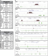
RNA-Seq and ChIP-Seq on DLX3-deficient bones and osteoblasts identify molecular targets of Dlx3 regulation of bone mass. RNA-Seq differential gene expression profiling was performed on femurs from 5 wk Dlx3+/+ and Dlx3OCN-cKO males. Selected genes differentially expressed in the metaphysis (a) and diaphysis (b) of Dlx3OCN-cKO compared with Dlx3+/+ mice genes are organized by genes involved in ECM and genes related to the osteoblastic differentiation and ossification processes. (c) ChIP-Seq analysis performed in SMAA-positive BMSCs shows DLX3 binding to bone-related genes differentially regulated in Dlx3OCN-cKO mice identified by RNA-Seq. Tracks for Dlx5, Dlx6, Sp7, Ibsp, Enpp1, Adamts18 and Tnfrsf11b (Opg) were visualized on UCSC genome browser with Refseq displayed for gene annotation and H3K4me3 track provided as reference
Figure 6
Figure 6
Dlx3 excision in calvarial osteoblasts increases expression of osteoblast markers. (a) Q-PCR of Dlx3 mRNA expression represented as fold-change in Adv-GFP and Adv-Cre-infected calvarial osteoblasts from Dlx3F/F neonates during proliferating (D4) and matrix maturation stages (D12). (b) Western blot of DLX3 and Lamin B in virus-infected calvarial osteoblasts at D12. (c) ALPL and Von Kossa stainings of virus-infected calvarial osteoblasts demonstrated increased ALPL activity and mineralization capacity in Adv-Cre-infected cells. (d) Q-PCR of mRNA expression represented as fold-change of osteoblast-related markers (Alpl, Ibsp, Ocn) and bone transcription factors (Runx2, Msx2, Dlx5). Data are presented as the mean±S.E.M. *P<0.05, **P<0.01, ***P<0.001
Figure 7
Figure 7
Increased occupancy of RUNX2 on the osteocalcin promoter in DLX3-deficient calvarial osteoblasts. (a) Diagram of the mouse osteocalcin promoter displaying relative binding sites and primer sites used for chromatin immunoprecipitation analysis. (b) Calvarial osteoblasts isolated from Dlx3F/F mice were infected with Adv-Cre or Adv-GFP and cultured in osteogenic media for 18 days. ChIP was then preformed on cleared cell lysates on the indicated day using ∼5 μg of RUNX2, DLX3, DLX5, or non-specific IgG antibody. Recovered DNA was then quantified by Q-PCR and normalized to input. Data are presented as the mean of three experiments±S.E.M. *P<0.05, **P<0.01, ***P<0.001

Comment in

References

    1. Cohen MM., Jr Perspectives on RUNX genes: an update. Am J Med Genet A. 2009;149A:2629–2646. - PubMed
    1. Jensen ED, Gopalakrishnan R, Westendorf JJ. Regulation of gene expression in osteoblasts. Biofactors. 2010;36:25–32. - PMC - PubMed
    1. Long F. Building strong bones: molecular regulation of the osteoblast lineage. Nat Rev Mol Cell Biol. 2012;13:27–38. - PubMed
    1. Depew MJ, Simpson CA, Morasso M, Rubenstein JL. Reassessing the Dlx code: the genetic regulation of branchial arch skeletal pattern and development. J Anat. 2005;207:501–561. - PMC - PubMed
    1. Panganiban G, Rubenstein JL. Developmental functions of the Distal-less/Dlx homeobox genes. Development. 2002;129:4371–4386. - PubMed

Publication types

Substances