. 2014 Jul 31;20(3):294-317.
doi: 10.5056/jnm14060.

The significance of interstitial cells in neurogastroenterology

Affiliations

The significance of interstitial cells in neurogastroenterology

Peter J Blair et al. J Neurogastroenterol Motil. .

Abstract

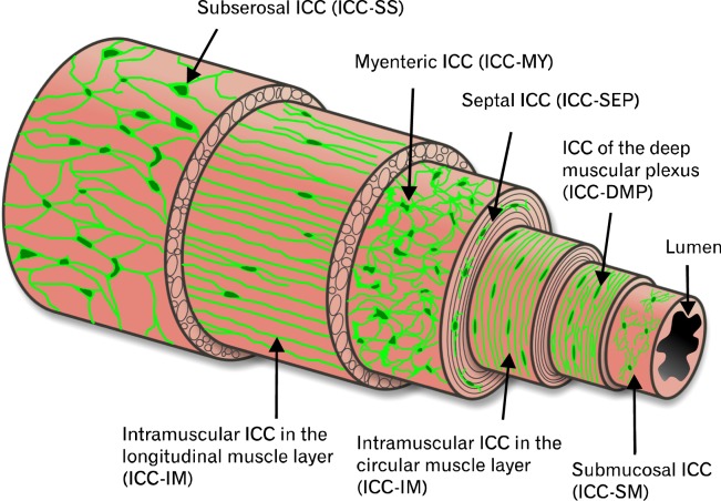
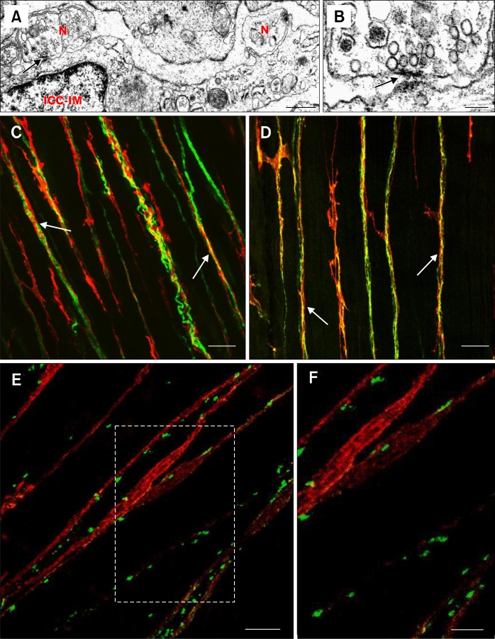
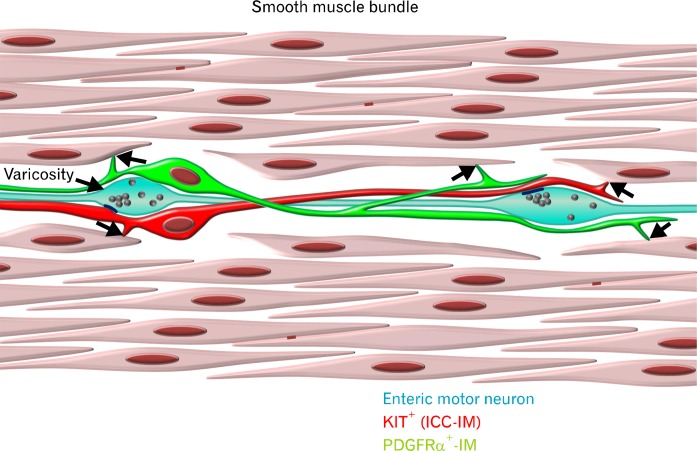
Smooth muscle layers of the gastrointestinal tract consist of a heterogeneous population of cells that include enteric neurons, several classes of interstitial cells of mesenchymal origin, a variety of immune cells and smooth muscle cells (SMCs). Over the last number of years the complexity of the interactions between these cell types has begun to emerge. For example, interstitial cells, consisting of both interstitial cells of Cajal (ICC) and platelet-derived growth factor receptor alpha-positive (PDGFRα(+)) cells generate pacemaker activity throughout the gastrointestinal (GI) tract and also transduce enteric motor nerve signals and mechanosensitivity to adjacent SMCs. ICC and PDGFRα(+) cells are electrically coupled to SMCs possibly via gap junctions forming a multicellular functional syncytium termed the SIP syncytium. Cells that make up the SIP syncytium are highly specialized containing unique receptors, ion channels and intracellular signaling pathways that regulate the excitability of GI muscles. The unique role of these cells in coordinating GI motility is evident by the altered motility patterns in animal models where interstitial cell networks are disrupted. Although considerable advances have been made in recent years on our understanding of the roles of these cells within the SIP syncytium, the full physiological functions of these cells and the consequences of their disruption in GI muscles have not been clearly defined. This review gives a synopsis of the history of interstitial cell discovery and highlights recent advances in structural, molecular expression and functional roles of these cells in the GI tract.

Keywords: Enteric nervous system, Receptor, platelet-derived growth factor alpha, SIP syncytium, Slow waves, TMEM16A protein, mouse.


Figures

Figure 1.
Figure 1.
Illustration depicting the various subpopulations of interstitial cells of Cajal (ICC) and their relative locations in the gut wall from several organs. Subserosal ICC (ICC-SS), located between the serosa and longitudinal muscle layer, are present in the small intestine and colon of various species. ICC-MY are located within the intermuscular plane between the circular and longitudinal layers throughout the gastrointestinal (GI) tract of all species studied. ICC-IM represent intramuscular ICC located within the circular and longitudinal muscle layers and are a prominent population of ICC in the stomach and colon. ICC-DMP represent a population of cells within the specialized deep muscular plexus region of the small intestine. In the small intestine of primates ICC-IM are also found within the circular muscle layer. Submucosal ICC (ICC-SM) are found on the inner aspect of the circular muscle layer and are prominent in the gastric antrum and colon.
Figure 2.
Figure 2.
Loss of pacemaker activity in mice with disrupted interstitial cells of Cajal (ICC) networks. (A) Wild type mice with normal slow waves (a) lose slow wave activity (b) in the small intestine after intraperitoneal injection with the KIT neutralizing antibody ACK2. (B) Slow waves present in wild type controls (a) are lost in small intestines of mice (b) that have a mutation in the KIT receptor (W/WV). Signaling via the KIT receptor (i.e., stem cell factor receptor) in ICC is essential for their survival. (C) Sl/Sld mice that have a mutation in stem cell factor, the ligand for the KIT receptor, lack slow waves in the small intestine (b) compared to wild type controls (a). Recordings in panels A, B and C were performed in the presence of L-type calcium channel blocker, nifedipine, in order to block muscle contraction and thus facilitate cell impalement. (D) Recordings from wild type and Sl/Sld mice in the absence of nifedipine: (a) Calcium action potentials are visible on the peaks of most regular slow waves in wild type mice. (b) In Sl/Sld mice irregular clusters of Ca2+ action potentials are observed in the absence of slow waves, demonstrating that the smooth muscle tissue is still capable of producing action potentials in the absence of ICC. Adapted from Torihashi et al (A), Ward et al (B) and Ward et al (C and D).
Figure 3.
Figure 3.
The intracellular mechanism underlying generation of pacemaker activity in interstitial cells of Cajal (ICC). (A) Stochastic release of Ca2+ from intracellular stores in ICC results in transient activation of anoctamin-1 (ANO1) channels (B), producing spontaneous transient inward currents (STICs) in primary pacemaker cells. The Na-K-Cl cotransporter (NKCC1) replenishes intracellular chloride ions. (C) The depolarization of ICC activates voltage-dependent, dihydropyridine-resistant Ca2+ channels (VDCC) in the plasma membrane (PM). Ca2+ entry causes additional Ca2+ release from IP3-depenent Ca2+ channels (IP3R1) and summation of ANO1 currents, resulting in slow wave generation. Slow waves depolarize nearby ICC (horizontal arrow) triggering activation of voltage-dependent Ca2+ channels in adjacent secondary pacemaker cells. Ca2+ entry in adjacent cells activates ANO1 channels and regenerates slow waves. Spread of events in this fashion occurs throughout ICC networks. Termination of slow waves occurs by reuptake of Ca2+ into the endoplasmic reticulum (ER) via the sarco/ER Ca2+-ATPase pump (SERCA).
Figure 4.
Figure 4.
Representative electrical slow wave from the human gastric antrum. Slow waves consist of several discrete phases. An initial upstroke phase (1) rises from a diastolic membrane potential of −70 mV (0). Phase 1 is thought to be due to release of intracellular calcium from the endoplasmic reticulum and activation of anoctamin-1 and voltage-dependent, dihydropyridine-resistant Ca2+ channels channels. The transient depolarization to −26 mV (2) is followed by a partial repolarization, likely due to activation of an A-type potassium conductance in smooth muscle cells, to a plateau phase (3) that is sustained for several seconds. Phase 3 is likely to be caused by a balance between inward and outward conductances (i.e., calcium ion entry, chloride ion efflux versus potassium ion efflux). The membrane potential then returns to the resting diastolic potential during phase 4.
Figure 5.
Figure 5.
Close associations and synaptic-like specializations between enteric neurons and interstitial cells of Cajal (ICC). (A) Transmission electron micrograph showing areas of increased electron density at sites where enteric neurons (N) and intramuscular ICC (ICC-IM) are closely apposed in the murine gastric antrum (arrow). Electron densifications were observed on both pre- and post-synaptic membranes. (B) The site indicated by the arrow in A is shown at higher magnification in B. The arrow in B also indicates areas of increased electron density as in A. (C and D) Double label immunohistochemistry images that show close structural relationships (arrows) between nerve fibers (green) and ICC (KIT; red) in the taenia coli of primate colon. (C) Close apposition between the pan-neuronal label, PGP9.5 (protein gene product 9.5) and ICC. (D) shows the relationship between inhibitory nNOS+ nerve fibers and ICC. (E) Double labeling of the pre-synaptic soluble N-ethylmaleimide-sensitive factor attachment protein receptor (SNARE) protein, synaptotagmin (green) and ICC (red) in murine fundus. The image demonstrates that pre-synaptic proteins are closely apposed to ICC, suggesting that ICC are likely the innervated post-synaptic cell. Area in (E) outlined by white box is shown at higher magnification in panel (F). Scale bars: A, 0.5 μm; B, 0.1 μm; C, 25 μm; D, 25 μm; E, 10 μm; and F, 5 μm. Adapted from Beckett et al (A, B, E and F) and Blair et al (C and D).
Figure 6.
Figure 6.
Diagrammatic representation of the SIP syncytium. The multicellular electrical syncytium consists of at least three distinct cell types. Kit+ interstitial cells of Cajal (ICC; red) and PDGFRα+ interstitial cells (green) form low-resistance gap junctions with each other and neighboring smooth muscle cells (SMCs; arrows). SMCs form gap junctions with each other (red lines). Enteric motor nerves (light blue) are closely apposed to both classes of interstitial cells and make synapse-like contacts with ICC (dark blue lines) at vesicle laden varicosities.

References

    1. Cajal SR. Sur les ganglions et plexus nerveux de l’intestin. CR Soc Biol. 1893;45:217–223.
    1. Cajal SR. Nuevas aplicaciones del metodo de coloracion De Golgi. Gaceta Medica Catalana. 1889;12:613–616.
    1. Cajal SR. El plexo de Auerbach de los batracios. Nota sobre el plexo de Auerbach de la rana. Trab Lab Histol Fac Med Barcelona. 1892:23–28.
    1. Cajal SR. Les nouvelles idées sur la structure du système nerveux. Paris: C. Reinwald & Co.; 1894.
    1. Cajal SR. Histologie du systeme nerveux de l’homme et des vertebres. Volume 2. Paris: Maloine; 1911. pp. 891–942.

LinkOut - more resources