

Clinical decision making with myocardial perfusion imaging in patients with known or suspected coronary artery disease
- PMID: 24948154
- PMCID: PMC4316685
- DOI: 10.1053/j.semnuclmed.2014.04.006
Clinical decision making with myocardial perfusion imaging in patients with known or suspected coronary artery disease
Abstract
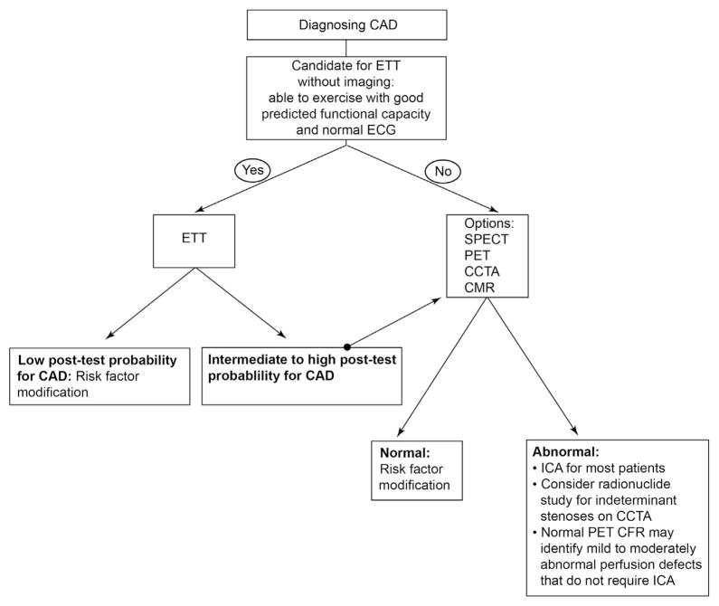
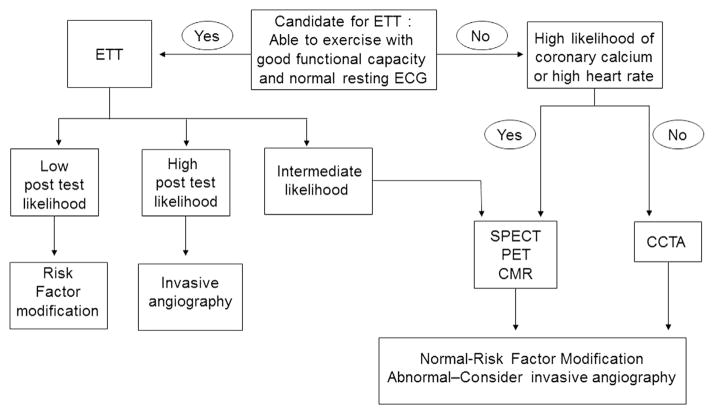
Myocardial perfusion imaging (MPI) to diagnose coronary artery disease (CAD) is best performed in patients with intermediate pretest likelihood of disease; unfortunately, pretest likelihood is often overestimated, resulting in the inappropriate use of perfusion imaging. A good functional capacity often predicts low risk, and MPI for diagnosing CAD should be reserved for individuals with poor exercise capacity, abnormal resting electrocardiography, or an intermediate or high probability of CAD. With respect to anatomy-based testing, coronary CT angiography has a good negative predictive value, but stenosis severity correlates poorly with ischemia. Therefore decision making with respect to revascularization may be limited when a purely noninvasive anatomical test is used. Regarding perfusion imaging, the diagnostic accuracies of SPECT, PET, and cardiac magnetic resonance are similar, though fewer studies are available with cardiac magnetic resonance. PET coronary flow reserve may offer a negative predictive value sufficiently high to exclude severe CAD such that patients with mild to moderate reversible perfusion defects can forego invasive angiography. In addition, combined anatomical and perfusion-based imaging may eventually offer a definitive evaluation for diagnosing CAD, even in higher risk patients. Any remarkable findings on single-photon emission computed tomography and PET MPI studies are valuable for prognostication. Furthermore, assessment of myocardial blood flow with PET is particularly powerful for prognostication as it reflects the end result of many processes that lead to atherosclerosis. Decision making with respect to revascularization is limited for cardiac MRI and PET MPI. In contrast, retrospective radionuclide studies have identified an ischemic threshold, but randomized trials are needed. In patients with at least moderately reduced left ventricular systolic function, viable myocardium as assessed by PET or MRI, appears to identify patients who benefit from revascularization, but well-executed randomized trials are lacking.
Copyright © 2014. Published by Elsevier Inc.
Figures
References
-
- Thomas JD. Myocardial contrast echocardiography perfusion imaging: Still waiting after all these years. J Am Coll Cardiol. 2013;62(15):1362–1364. - PubMed
-
- Rozanski A, Gransar H, Hayes SW, et al. Temporal trends in the frequency of inducible myocardial ischemia during cardiac stress testing: 1991 to 2009. J Am Coll Cardiol. 2013;61(10):1054–1065. - PubMed
-
- Shaw LJ, Mieres JH, Hendel RH, et al. Comparative effectiveness of exercise electrocardiography with or without myocardial perfusion single photon emission computed tomography in women with suspected coronary artery disease: Results from the what is the optimal method for ischemia evaluation in women (WOMEN) trial. Circulation. 2011;124 (11):1239–1249. - PubMed
-
- Christman MP, Marcio Sommer Bittencourt MD, Edward Hulten MM, et al. The yield of downstream tests after exercise treadmill testing: A prospective cohort study. J Am Coll Cardiol. 2014;63(13):1264–1274. http://dx.doi.org/10.1016/j.jacc.2013.11.052. [Epub ahead of print] - DOI - PMC - PubMed
Publication types
MeSH terms
Grants and funding
LinkOut - more resources
Full Text Sources
Other Literature Sources
Medical
Miscellaneous
