. 2014 Jun 19:11:111.
doi: 10.1186/1742-2094-11-111.

Human APOE4 increases microglia reactivity at Aβ plaques in a mouse model of Aβ deposition

Affiliations

Human APOE4 increases microglia reactivity at Aβ plaques in a mouse model of Aβ deposition

Gustavo A Rodriguez et al. J Neuroinflammation. .

Abstract

Background: Having the apolipoprotein E4 (APOE-ϵ4) allele is the strongest genetic risk factor for the development of Alzheimer's disease (AD). Accumulation of amyloid beta (Aβ) in the brain is influenced by APOE genotype. Transgenic mice co-expressing five familial AD mutations (5xFAD) in the presence of human APOE alleles (ϵ2, ϵ3 or ϵ4) exhibit APOE genotype-specific differences in early Aβ accumulation, suggesting an interaction between APOE and AD pathology. Whether APOE genotype affects Aβ-plaque-associated neuroinflammation remains unclear. In the current study, we address the role of APOE genotype on Aβ-associated microglial reactivity in the EFAD transgenic mouse model.

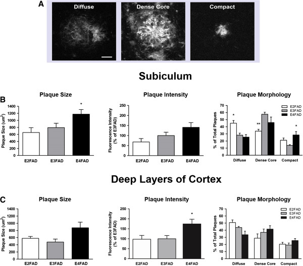
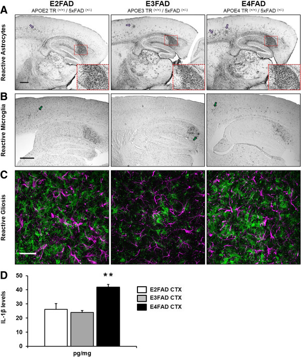
Methods: We analyzed Aβ-induced glial activation in the brains of 6-month-old EFAD transgenic mice (E2FAD, E3FAD and E4FAD). Region-specific morphological profiles of Aβ plaques in EFAD brain sections were compared using immunofluorescence staining. We then determined the degree of glial activation in sites of Aβ deposition while comparing levels of the inflammatory cytokine Interleukin-1β (IL-1β) by ELISA. Finally, we quantified parameters of Aβ-associated microglial reactivity using double-stained EFAD brain sections.

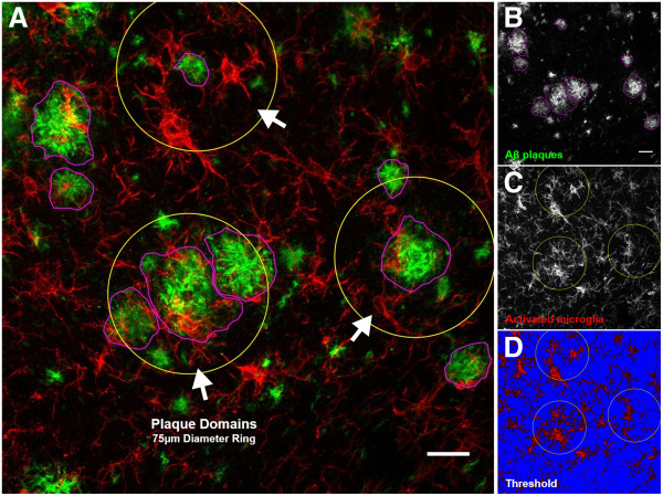
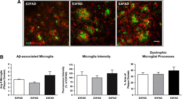
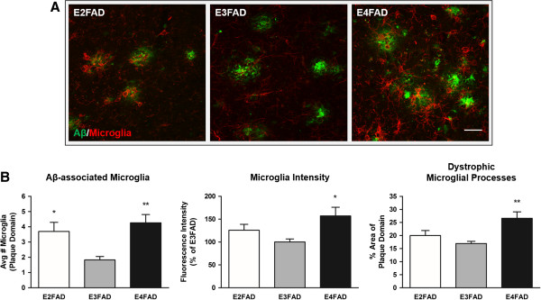
Results: Characterization of Aβ plaques revealed there were larger and more intensely stained plaques in E4FAD mice relative to E2FAD and E3FAD mice. E4FAD mice also had a greater percentage of compact plaques in the subiculum than E3FAD mice. Reactive microglia and dystrophic astrocytes were prominent in EFAD brains, and primarily localized to two sites of significant Aβ deposition: the subiculum and deep layers of the cortex. Cortical levels of IL-1β were nearly twofold greater in E4FAD mice relative to E3FAD mice. To control for differences in levels of Aβ in the different EFAD mice, we analyzed the microglia within domains of specific Aβ deposits. Morphometric analyses revealed increased measures of microglial reactivity in E4FAD mice, including greater dystrophy, increased fluorescence intensity and a higher density of reactive cells surrounding cortical plaques, than in E3FAD mice.

Conclusions: In addition to altering morphological profiles of Aβ deposition, APOE genotype influences Aβ-induced glial activation in the adult EFAD cortex. These data support a role for APOE in modulating Aβ-induced neuroinflammatory responses in AD progression, and support the use of EFAD mice as a suitable model for mechanistic studies of Aβ-associated neuroinflammation.


Figures

Figure 1
Figure 1
APOE genotype affects Aβ plaque morphology in EFAD subiculum and deep layers of cortex. Morphologically distinct Aβ plaques were visualized in the subiculum and deep layers of the cortex of 6-month-old EFAD mice using immunofluorescence staining with Aβ antibody MOAB2. (A) Heterogeneous plaque types were evident in EFAD brains and grouped into three categories: diffuse, dense core and compact. Scale bar: 12 μm. (B) Subiculum. Left: E4FAD mice exhibited larger plaques on average than E3FAD mice. Center: Fluorescence intensity of plaques did not differ by APOE genotype. Right: E2FAD mice exhibited a greater percentage of diffuse plaques than E3FAD or E4FAD mice, and a lower percentage of dense core plaques than E3FAD mice. Interestingly, E4FAD mice exhibited an increased percentage of compact plaques in the subiculum. (C) Deep layers of the cortex. Left: Plaque size did not differ by APOE genotype. Center: Fluorescence intensity of deep cortical plaques was highest in E4FAD mice. Right: E2FAD mice had a greater percentage of diffuse plaques in this region than E4FAD mice. No differences were detected between APOE genotype for dense core or compact plaques. One-way ANOVA, * P < 0.05. Two-way ANOVA, * P < 0.05, ** P < 0.01.
Figure 2
Figure 2
Glial activation is increased in the EFAD subiculum and cortex, while E4FAD mice exhibit elevated IL-1β levels. Astrocytes and microglia were visualized in sagittal brain sections of 6-month-old EFAD mice using immunohistochemistry. Initial DAB staining revealed prominent gliosis in two regions of the brain: the subiculum and deep layers of the cortex. (A) Extensive GFAP-immunoperoxidase staining was evident in the subiculum of all EFAD mice. In addition, dystrophic astrocytes could be seen throughout the deep layers, but not in superficial layers, of the EFAD cortex. Insets: 20× magnification of the subiculum (dashed red box). Magenta arrows: Clusters of dystrophic astrocytes in the cortex. Scale bar: 500 μm. (B) Activated microglial cells were clearly visible in the subiculum of all EFAD mice and were present in deep cortical layers as well. E4FAD sections exhibited more activated microglia in the deep cortex than E2FAD and E3FAD sections. Green arrows: Activated microglia. Scale bar: 500 μm. (C) Double immunofluorescence staining for GFAP (magenta) and Iba1 (green) confirmed region-specific glial activation in EFAD brains. Representative images of EFAD subicula are shown. Scale bar: 30 μm. (D) Levels of the pro-inflammatory cytokine IL-1β were measured using ELISA in 6-month-old EFAD cortex (CTX) extracts. E4FAD sections exhibited significantly higher levels of IL-1β than E3FAD sections. One-way ANOVA, ** P < 0.01.
Figure 3
Figure 3
Confocal microscopy analysis of Aβ-associated microglial activation in EFAD mice. Sagittal brain sections from 6-month-old EFAD mice were analyzed for Aβ deposition and associated microglial activation in two regions of interest, the subiculum and deep layers of the cortex. Reactive microglial cells within the vicinity of Aβ plaques consistently exhibited an amoeboid-like shape with dystrophic processes. (A) Two-dimensional projection image (approximately 20 μm, z-axis) of a E3FAD subiculum double stained for Aβ1–42 (MOAB2) and activated microglia (Iba1). Image overlays depict metrics used to characterize and quantify Aβ deposition and associated microglial activation. White arrows: Examples of plaque domains. (B) Grayscale image of Aβ plaques chosen for analysis. Plaque types, size of plaques and MOAB2 fluorescence intensity were quantified. Magenta: Traced plaques. (C) Grayscale image of activated microglia with plaque domain overlays. The number of microglia cell bodies within plaque domains were collected and quantified along with Iba1 fluorescence intensity. Yellow ring: Plaque domains (4.42 mm2). (D) Thresholding was performed within plaque domains to quantify the percentage area occupied by microglia cell bodies and processes. Representative minimum threshold value of 35 (0 to 255 brightness scale) depicted. Blue: Background. Scale bars: 20 μm.
Figure 4
Figure 4
Aβ deposition in the subiculum is associated with high levels of microglial reactivity. Double immunofluorescence staining of 6-month-old EFAD subicula revealed significant Aβ deposition and elevated microglial activation in each group. (A) Representative images of fluorescence staining in EFAD subiculum. Aβ-associated microglial activation was assessed for each APOE genotype (n = 4 or 5 mice/APOE genotype). Scale bar: 30 μm. (B) Microglial density, fluorescence intensity and percentage area of the plaque domain did not differ between APOE genotypes. Within the subiculum, the reactive microglia in each group occupies a large percentage of the plaque domains relative to the deep cortex.
Figure 5
Figure 5
APOE4 is associated with increased microglial reactivity to Aβ in deep layers of the cortex. Morphological profiles of reactive microglia within deep cortical Aβ plaque domains were elevated in E4FAD mice. (A) Representative double-stained images of Aβ plaques and microglial activation in deep layers of EFAD cortices (n = 4 or 5 mice/APOE genotype). Scale bar: 30 μm. (B) Left: E4FAD and E2FAD mice exhibited increased reactive microglial density within Aβ plaque domains. Center: Fluorescence intensity of the microglia was elevated in E4FAD mice relative to E3FAD mice. Right: E4FAD microglia occupied a larger percentage of plaque domains than E3FAD microglia. One-way ANOVA, * P < 0.05, ** P < 0.01.

References

    1. Selkoe DJ. Resolving controversies on the path to Alzheimer's therapeutics. Nat Med. 2011;17:1060–1065. - PubMed
    1. Selkoe DJ. Alzheimer's disease. Cold Spring Harb Perspect Biol. 2011;3(7):1–16. - PMC - PubMed
    1. Braak H, Braak E. Staging of Alzheimer's disease-related neurofibrillary changes. Neurobiol Aging. 1995;16:271–278. discussion 278–284. - PubMed
    1. Hardy J, Selkoe DJ. The amyloid hypothesis of Alzheimer's disease: progress and problems on the road to therapeutics. Science. 2002;297:353–356. - PubMed
    1. Amor S, Puentes F, Baker D, van der Valk P. Inflammation in neurodegenerative diseases. Immunology. 2010;129:154–169. - PMC - PubMed

Publication types

MeSH terms