

Two-site reproducibility of cerebellar and brainstem neurochemical profiles with short-echo, single-voxel MRS at 3T
- PMID: 24948590
- PMCID: PMC4272339
- DOI: 10.1002/mrm.25295
Two-site reproducibility of cerebellar and brainstem neurochemical profiles with short-echo, single-voxel MRS at 3T
Abstract
Purpose: To determine whether neurochemical concentrations obtained at two MRI sites using clinical 3T scanners can be pooled when a highly optimized, nonvendor short-echo, single-voxel proton MRS pulse sequence is used in conjunction with identical calibration and quantification procedures.
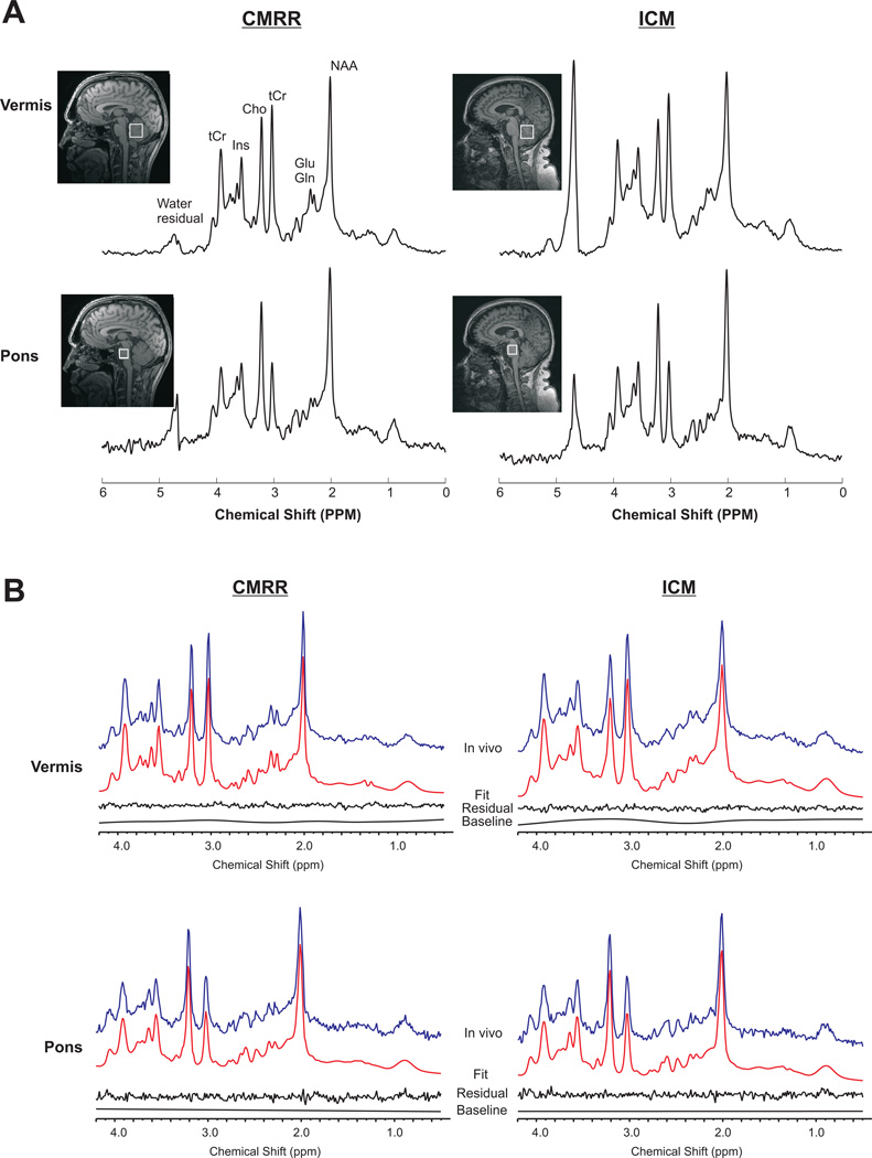
Methods: A modified semi-LASER sequence (TE = 28 ms) was used to acquire spectra from two brain regions (cerebellar vermis and pons) on two Siemens 3T scanners using the same B0 and B1 calibration protocols from two different cohorts of healthy volunteers (N = 24-33 per site) matched for age and body mass index. Spectra were quantified with LCModel using water scaling.
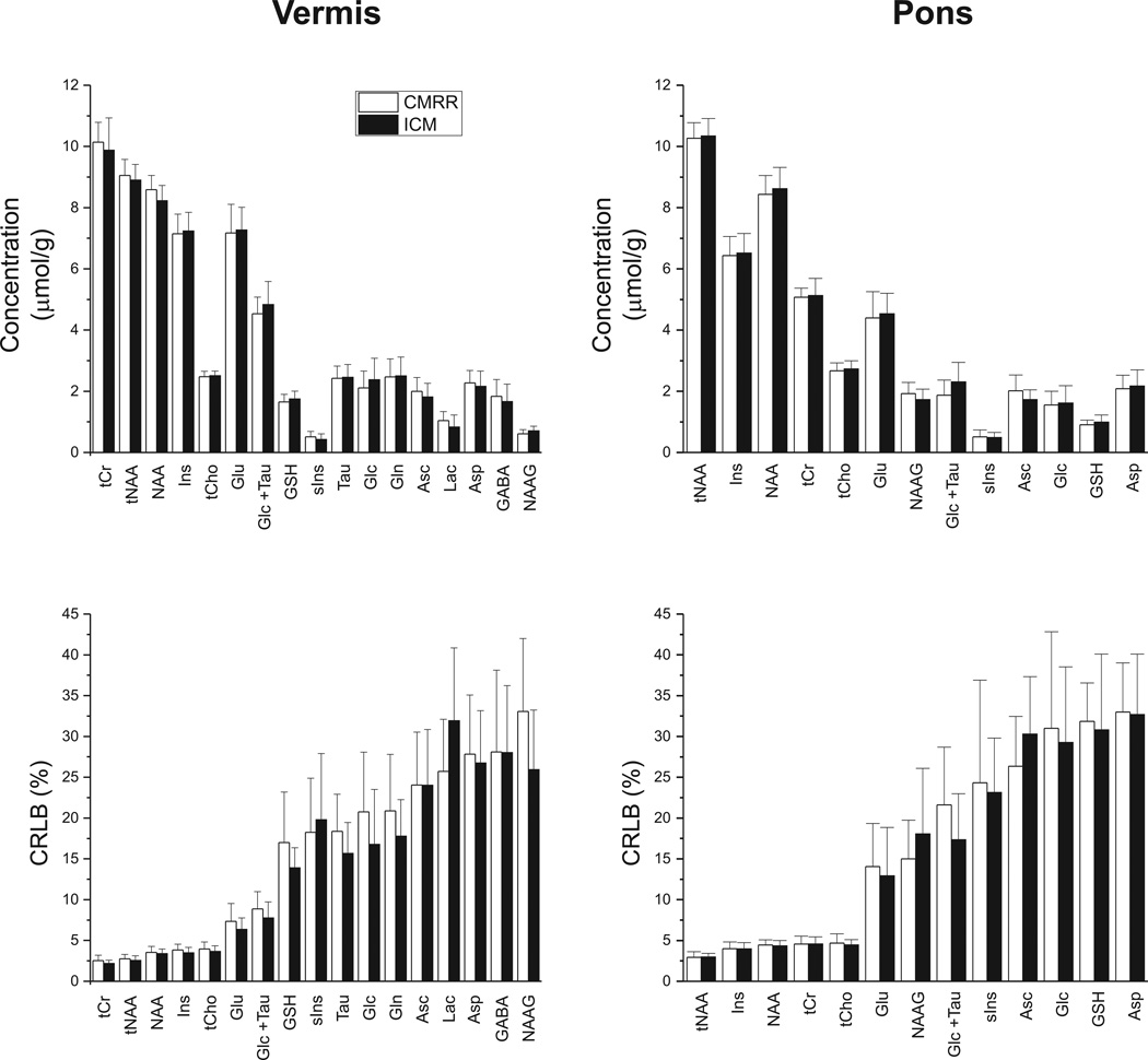
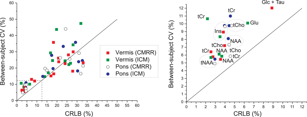
Results: The spectral quality was very consistent between the two sites and allowed reliable quantification of at least 13 metabolites in the vermis and pons compared with 3-5 metabolites in prior multisite magnetic resonance spectroscopy trials using vendor-provided sequences. The neurochemical profiles were nearly identical at the two sites and showed the feasibility to detect interindividual differences in the healthy brain.
Conclusion: Highly reproducible neurochemical profiles can be obtained on different clinical 3T scanners at different sites, provided that the same, optimized acquisition and analysis techniques are used. This will allow pooling of multisite data in clinical studies, which is particularly critical for rare neurological diseases.
Keywords: 3 Tesla; multi-site; reproducibility; spectroscopy.
© 2014 Wiley Periodicals, Inc.
Figures
References
-
- Öz G, Alger J, Barker P, Bartha R, Bizzi A, Boesch C, Bolan P, Brindle K, Cudalbu C, Dincer A, Dydak U, Emir U, Frahm J, Gonzalez R, Gruber S, Gruetter R, Gupta R, Heerschap A, Henning A, Hetherington H, Howe F, Huppi P, Hurd R, Kantarci K, Klomp D, Kreis R, Kruiskamp M, Leach M, Lin A, Luijten P, Marjanska M, Maudsley A, Meyerhoff D, Mountford C, Nelson S, Ozduman K, Necmettin P, Pan J, Peet A, Poptani H, Posse S, Pouwels P, Ratai E, Ross B, Scheenen T, Schuster C, Soher B, Tkac I, Vigneron D, Kauppinen R. Clinical Proton MR Spectroscopy in Central Nervous System Disorders: The MRS Consensus Group. Radiology. 2014;270(3):658–679. - PMC - PubMed
-
- Wattjes MP. Structural MRI. International Psychogeriatrics. 2011;23(Supplement S2):S13–S24. - PubMed
-
- Hentschel F, Kreis M, Damian M, Krumm B, Frolich L. The clinical utility of structural neuroimaging with MRI for diagnosis and differential diagnosis of dementia: a memory clinic study. Int J Geriat Psychiatry. 2005;20(7):645–650. - PubMed
-
- Komoroski RA, Kotrla KJ, Lemen L, Lindquist D, Diaz P, Foundas A. Brain metabolite concentration ratios in vivo: multisite reproducibility by single-voxel 1H MR spectroscopy. Magn Reson Imaging. 2004;22(5):721–725. - PubMed
-
- Traber F, Block W, Freymann N, Gur O, Kucinski T, Hammen T, Ende G, Pilatus U, Hampel H, Schild H, Heun R, Jessen F. A multicenter reproducibility study of single-voxel 1H-MRS of the medial temporal lobe. European Radiology. 2006;16:1096–1103. - PubMed
Publication types
MeSH terms
Grants and funding
LinkOut - more resources
Full Text Sources
Other Literature Sources
