

Primary stability of self-drilling and self-tapping mini-implant in tibia of diabetes-induced rabbits
- PMID: 24949013
- PMCID: PMC4037616
- DOI: 10.1155/2014/429359
Primary stability of self-drilling and self-tapping mini-implant in tibia of diabetes-induced rabbits
Abstract
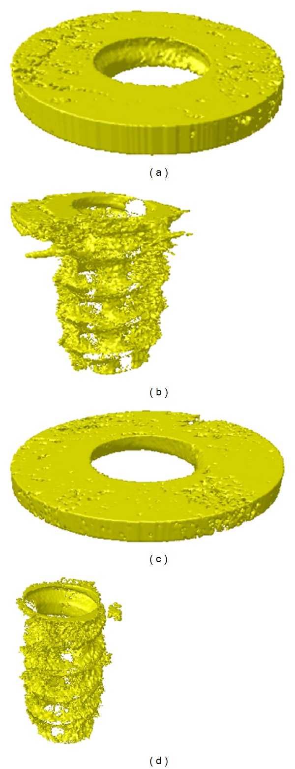
Objective. This study aimed to evaluate effects of type 1 diabetes mellitus and mini-implant placement method on the primary stability of mini-implants by comparing mechanical stability and microstructural/histological differences. Methods. After 4 weeks of diabetic induction, 48 mini-implants (24 self-tapping and 24 self-drilling implants) were placed on the tibia of 6 diabetic and 6 normal rabbits. After 4 weeks, the rabbits were sacrificed. Insertion torque, removal torque, insertion energy, and removal energy were measured with a surgical engine on 8 rabbits. Remaining 4 rabbits were analyzed by microcomputed tomography (micro-CT) and bone histomorphometry. Results. Total insertion energy was higher in self-drilling groups than self-tapping groups in both control and diabetic groups. Diabetic groups had more trabecular separation in bone marrow than the control groups in both SD and ST groups. Micro-CT analysis showed deterioration of bone quality in tibia especially in bone marrow of diabetic rabbits. However, there was no statistically significant correlation between self-drilling and self-tapping group for the remaining measurements in both control and diabetic groups. Conclusions. Type 1 diabetes mellitus and placement method of mini-implant did not affect primary stability of mini-implants.
Figures


References
-
- Miyawaki S, Koyama I, Inoue M, Mishima K, Sugahara T, Takano-Yamamoto T. Factors associated with the stability of titanium screws placed in the posterior region for orthodontic anchorage. The American Journal of Orthodontics and Dentofacial Orthopedics. 2003;124(4):373–378. - PubMed
-
- Cheng SJ, Tseng IY, Lee JJ, Kok SH. A prospective study of the risk factors associated with failure of mini-implants used for orthodontic anchorage. International Journal of Oral and Maxillofacial Implants. 2004;19(1):100–106. - PubMed
-
- Schätzle M, Männchen R, Zwahlen M, Lang NP. Survival and failure rates of orthodontic temporary anchorage devices: a systematic review. Clinical Oral Implants Research. 2009;20(12):1351–1359. - PubMed
-
- Heidemann W, Terheyden H, Louis Gerlach K. Analysis of the osseous/metal interface of drill free screws and self-tapping screws. Journal of Cranio-Maxillofacial Surgery. 2001;29(2):69–74. - PubMed
-
- Sowden D, Schmitz JP. AO self-drilling and self-tapping screws in rat calvarial bone: an ultrastructural study of the implant interface. Journal of Oral and Maxillofacial Surgery. 2002;60(3):294–299. - PubMed
LinkOut - more resources
Full Text Sources
Other Literature Sources
