

Alveolar antral artery isolation during sinus lift procedure with the double window technique
- PMID: 24949106
- PMCID: PMC4062959
- DOI: 10.2174/1874210601408010095
Alveolar antral artery isolation during sinus lift procedure with the double window technique
Abstract
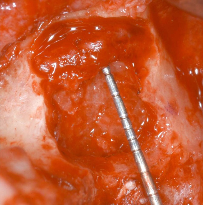
The sinus lift technique, introduced in 1976 by Tatum and subsequently described by Boyne in 1980, is nowadays considered a safe and reliable procedure for the rehabilitation of the atrophic upper posterior maxilla. The alveolar antral artery (AAA) is anastomoses between the posterior superior alveolar artery (PSAA) and the infraorbital artery (IOA) and may be present in the sinusal antrostomy. The haemorrhage of this vascular bundle represents the second intra-operatory complication in term of frequency during sinus lift procedure. Purpose of this study was to illustrate and describe a new technique allowing the AAA isolation during sinus lift procedure in cases in which the artery is clearly present inside the surgical area, detectable through CT scan exam. Presence, course and possible identification of the alveolar antral artery are also discussed, according to the studies present in the literature.
Keywords: Alveolar antral artery; sinus lift complications..
Figures


References
-
- Solar P, Geyerhofer U, Traxler H, Windisch A, Ulm C, Watzek G. Blood supply to the maxillary sinus relevant to sinus floor elevation procedures. Clin Oral Implants Res. 1999;10:34–44. - PubMed
-
- Elian N, Wallace S, Cho SC, Jalbout ZN, Froum S. Distribution of the maxillary artery as it relates to sinus floor augmentation. Int J Oral Maxillofac Implants. 2005;20:784–7. - PubMed
-
- Tatum H Jr. Maxillary and sinus implant reconstructions. Dent Clin North Am. 1986;30:207–29. - PubMed
-
- Chanovaz M. Maxillary sinus: anatomy. physioogy.surgery., and bone grafting related to implantology--eleven years of surgical experience 1979-1990). J Oral Implantol . 1990; 16:199–209. - PubMed
-
- Boyne PJ, James RA. Grafting of the maxillary sinus floor with autogenous marrow and bone. J Oral Surg. 1980;38:613–6. - PubMed
LinkOut - more resources
Full Text Sources
Other Literature Sources