

Lung ultrasound imaging in avian influenza A (H7N9) respiratory failure
- PMID: 24949191
- PMCID: PMC4051407
- DOI: 10.1186/2036-7902-6-6
Lung ultrasound imaging in avian influenza A (H7N9) respiratory failure
Abstract
Background: Lung ultrasound has been shown to identify in real-time, various pathologies of the lung such as pneumonia, viral pneumonia, and acute respiratory distress syndrome (ARDS). Lung ultrasound maybe a first-line alternative to chest X-ray and CT scan in critically ill patients with respiratory failure. We describe the use of lung ultrasound imaging and findings in two cases of severe respiratory failure from avian influenza A (H7N9) infection.
Methods: Serial lung ultrasound images and video from two cases of H7N9 respiratory failure requiring mechanical ventilation and extracorporeal membrane oxygenation in a tertiary care intensive care unit were analyzed for characteristic lung ultrasound findings described previously for respiratory failure and infection. These findings were followed serially, correlated with clinical course and chest X-ray.
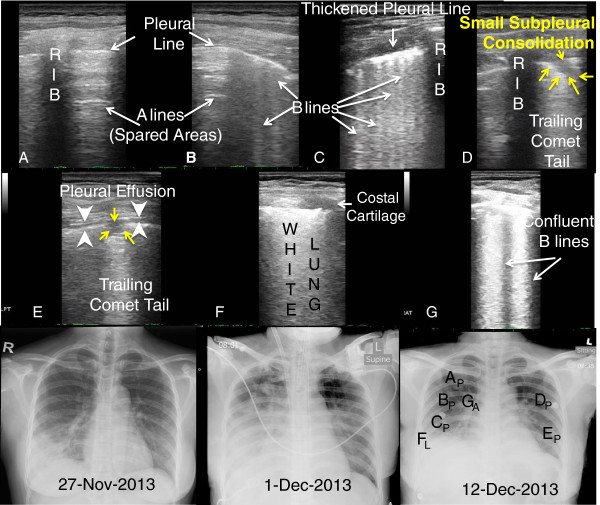
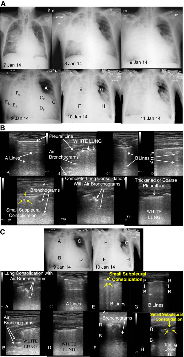
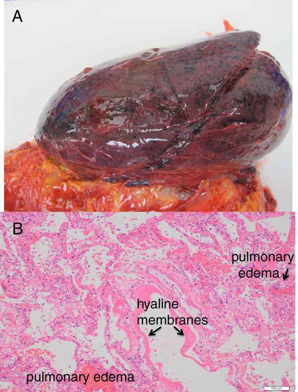
Results: IN BOTH PATIENTS, CHARACTERISTIC LUNG ULTRASOUND FINDINGS HAVE BEEN OBSERVED AS PREVIOUSLY DESCRIBED IN VIRAL PULMONARY INFECTIONS: subpleural consolidations associated or not with local pleural effusion. In addition, numerous, confluent, or coalescing B-lines leading to 'white lung' with corresponding pleural line thickening are associated with ARDS. Extension or reduction of lesions observed with ultrasound was also correlated respectively with clinical worsening or improvement. Coexisting consolidated pneumonia with sonographic air bronchograms was noted in one patient who did not survive.
Conclusions: Clinicians with access to point-of-care ultrasonography may use these findings as an alternative to chest X-ray or CT scan. Lung ultrasound imaging may assist in the efficient allocation of intensive care for patients with respiratory failure from viral pulmonary infections, especially in resource scarce settings or situations such as future respiratory virus outbreaks or pandemics.
Keywords: Critical care medicine; Emergency medicine; H7N9; Influenza A virus; Lung ultrasound; Pandemics; Point-of-care; Respiratory failure; Ultrasonography; Viral pneumonia.
Figures
References
-
- Gao HN, Lu HZ, Cao B, Du B, Shang H, Gan JH, Lu SH, Yang YD, Fang Q, Shen YZ, Xi XM, Gu Q, Zhou XM, Qu HP, Yan Z, Li FM, Zhao W, Gao ZC, Wang GF, Ruan LX, Wang WH, Ye J, Cao HF, Li XW, Zhang WH, Fang XC, He J, Liang WF, Xie J, Zeng M. et al.Clinical findings in 111 cases of influenza A (H7N9) virus infection. N Engl J Med. 2013;6(24):2277–2285. doi: 10.1056/NEJMoa1305584. - DOI - PubMed
-
- Li Q, Zhou L, Zhou M, Chen Z, Li F, Wu H, Xiang N, Chen E, Tang F, Wang D, Meng L, Hong Z, Tu W, Cao Y, Li L, Ding F, Liu B, Wang M, Xie R, Gao R, Li X, Bai T, Zou S, He J, Hu J, Xu Y, Chai C, Wang S, Gao Y, Jin L. et al.Epidemiology of human infections with avian influenza A (H7N9) virus in China. N Engl J Med. 2014;6(6):520–532. doi: 10.1056/NEJMoa1304617. doi:10.1056/NEJMoa1304617. Epub 2013 Apr 24. - DOI - PMC - PubMed
-
- Volpicelli G, Frascisco M. Sonographic detection of radio-occult interstitial lung involvement in measles pneumonia. Am J Emerg Med. 2009;6(1):128.e1–128.e3. - PubMed
LinkOut - more resources
Full Text Sources
Other Literature Sources
Research Materials
