

Intraductal papillary neoplasms of the bile duct
- PMID: 24949206
- PMCID: PMC4052179
- DOI: 10.1155/2014/459091
Intraductal papillary neoplasms of the bile duct
Abstract
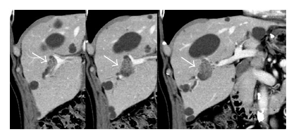
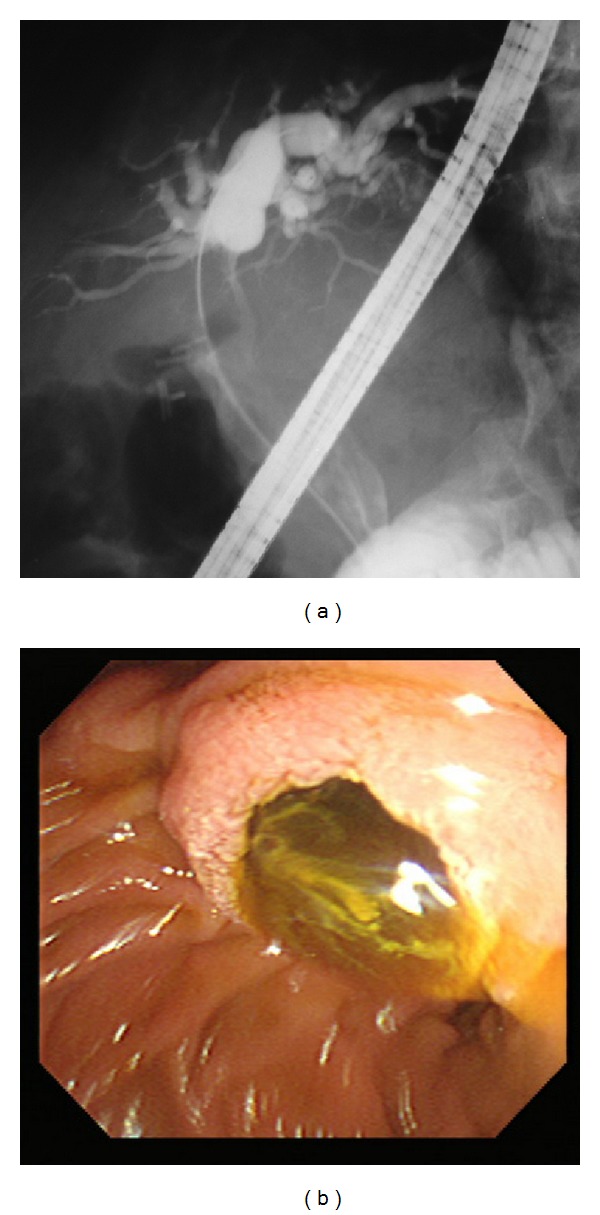
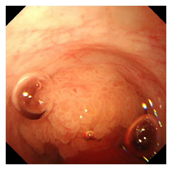
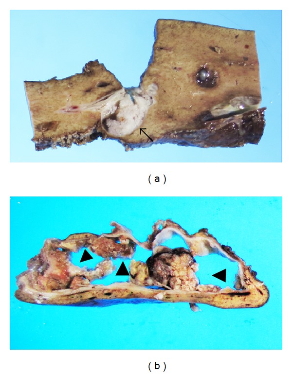
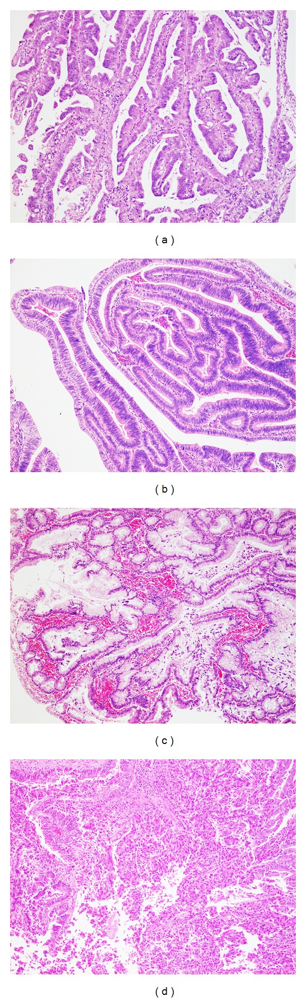
Intraductal papillary neoplasm of the bile duct (IPNB) is a rare variant of bile duct tumors characterized by papillary growth within the bile duct lumen and is regarded as a biliary counterpart of intraductal papillary mucinous neoplasm of the pancreas. IPNBs display a spectrum of premalignant lesion towards invasive cholangiocarcinoma. The most common radiologic findings for IPNB are bile duct dilatation and intraductal masses. The major treatment of IPNB is surgical resection. Ultrasonography, computed tomography, magnetic resonance image, and cholangiography are usually performed to assess tumor location and extension. Cholangioscopy can confirm the histology and assess the extent of the tumor including superficial spreading along the biliary epithelium. However, pathologic diagnosis by preoperative biopsy cannot always reflect the maximum degree of atypia, because IPNBs are often composed of varying degrees of cytoarchitectural atypia. IPNBs are microscopically classified into four epithelial subtypes, such as pancreatobiliary, intestinal, gastric, and oncocytic types. Most cases of IPNB are IPN with high-grade intraepithelial neoplasia or with an associated invasive carcinoma. The histologic types of invasive lesions are either tubular adenocarcinoma or mucinous carcinoma. Although several authors have investigated molecular genetic changes during the development and progression of IPNB, these are still poorly characterized and controversial.
Figures
Similar articles
-
Intraductal papillary neoplasm of the bile duct.World J Gastroenterol. 2013 Dec 14;19(46):8595-604. doi: 10.3748/wjg.v19.i46.8595. World J Gastroenterol. 2013. PMID: 24379576 Free PMC article. Review.
-
Intraductal Papillary Neoplasm of Bile Duct: Updated Clinicopathological Characteristics and Molecular and Genetic Alterations.J Clin Med. 2020 Dec 9;9(12):3991. doi: 10.3390/jcm9123991. J Clin Med. 2020. PMID: 33317146 Free PMC article. Review.
-
Intraductal papillary neoplasm of the bile duct: a biliary equivalent to intraductal papillary mucinous neoplasm of the pancreas?Hepatology. 2012 Oct;56(4):1352-60. doi: 10.1002/hep.25786. Epub 2012 Aug 27. Hepatology. 2012. PMID: 22504729
-
Clinicopathological characterization of so-called "cholangiocarcinoma with intraductal papillary growth" with respect to "intraductal papillary neoplasm of bile duct (IPNB)".Int J Clin Exp Pathol. 2014 May 15;7(6):3112-22. eCollection 2014. Int J Clin Exp Pathol. 2014. PMID: 25031730 Free PMC article.
-
Characterization of Intraductal Papillary Neoplasm of the Bile Duct with Respect to the Histopathologic Similarities to Pancreatic Intraductal Papillary Mucinous Neoplasm.Gut Liver. 2019 Nov 15;13(6):617-627. doi: 10.5009/gnl18476. Gut Liver. 2019. PMID: 30982236 Free PMC article. Review.
Cited by
-
Intraductal papillary neoplasm of the bile duct: diagnostic value of MRI features in differentiating pathologic subclassifications-type 1 versus type 2.Eur Radiol. 2024 Jul;34(7):4674-4685. doi: 10.1007/s00330-023-10491-9. Epub 2023 Dec 19. Eur Radiol. 2024. PMID: 38114846
-
Co-occurrence of IPMN and malignant IPNB complicated by a pancreatobiliary fistula: A case report and review of the literature.World J Clin Cases. 2019 Jan 6;7(1):102-108. doi: 10.12998/wjcc.v7.i1.102. World J Clin Cases. 2019. PMID: 30637259 Free PMC article.
-
Liver and Pancreas: Do Similar Embryonic Development and Tissue Organization Lead to Similar Mechanisms of Tumorigenesis?Gene Expr. 2018 Aug 22;18(3):149-155. doi: 10.3727/105221618X15216414278706. Epub 2018 Mar 26. Gene Expr. 2018. PMID: 29580319 Free PMC article. Review.
-
A rare case of extrahepatic biliary mucinous cystic neoplasm in a middle-aged woman: A case report.Mol Clin Oncol. 2021 Oct;15(4):196. doi: 10.3892/mco.2021.2358. Epub 2021 Jul 29. Mol Clin Oncol. 2021. PMID: 34462652 Free PMC article.
-
Intraductal Papillary Neoplasm of the Bile Duct: A Rare Case of Intrahepatic Space-Occupying Lesion.Cureus. 2021 Feb 1;13(2):e13063. doi: 10.7759/cureus.13063. Cureus. 2021. PMID: 33680605 Free PMC article.
References
-
- Sakamoto E, Nimura Y, Hayakawa N, et al. Clinicopathological studies of mucin-producing cholangiocarcinoma. Journal of Hepato-Biliary-Pancreatic Surgery. 1997;4(2):157–162.
-
- Shibahara H, Tamada S, Goto M, et al. Prognostic features of mucin-producing bile duct tumors. Two histopathologic categories as counterparts of pancreatic intraductal papillary-mucinous neoplasms. American Journal of Surgical Pathology. 2004;28(3):327–338. - PubMed
-
- Kuo C, Changchien C, Wu K, et al. Mucin-producing cholangiocarcinoma: clinical experience of 24 cases in 16 years. Scandinavian Journal of Gastroenterology. 2005;40(4):455–459. - PubMed
-
- Kim HJ, Kim MH, Lee SK, et al. Mucin-hypersecreting bile duct tumor characterized by a striking homology with an intraductal papillary mucinous tumor (IPMT) of the pancreas. Endoscopy. 2000;32(5):389–393. - PubMed
Publication types
LinkOut - more resources
Full Text Sources
Other Literature Sources
