

Atypical teratoid/rhabdoid tumor in sellar turcica in an adult: A case report and review of the literature
- PMID: 24949218
- PMCID: PMC4061576
- DOI: 10.4103/2152-7806.133105
Atypical teratoid/rhabdoid tumor in sellar turcica in an adult: A case report and review of the literature
Abstract
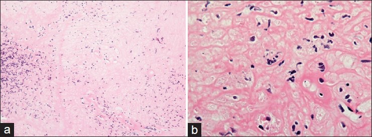
Background: Atypical teratoid/rhabdoid tumor (AT/RT) is a rare central nervous system tumor composed of primitive rhabdoid cells that may differentiate along neuroectodermal, mesenchymal and epithelial lineages. AT/RT in adults is rare but not completely exceptional. It generally arises from the posterior fossa of infants, but the broad majority of the reported AT/RT in adults manifested supratentorially with the exception of four cases that arose in the cerebellum and two that arose in the spinal cord.
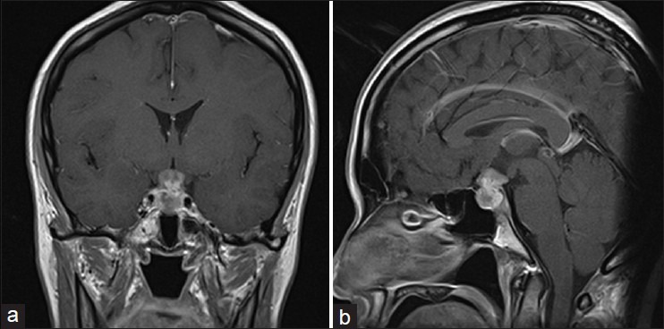
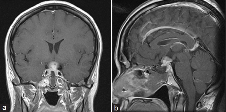
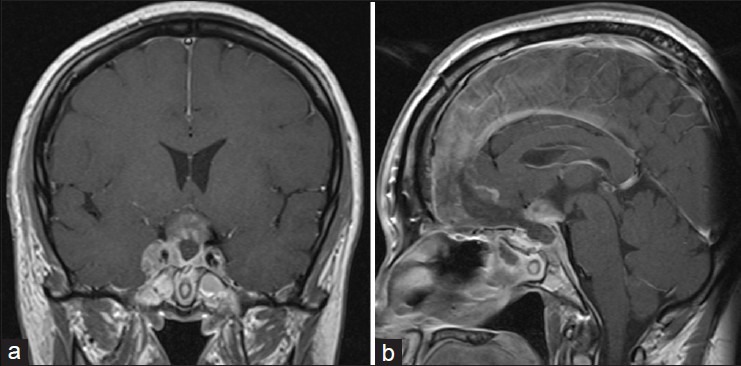
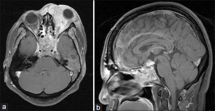
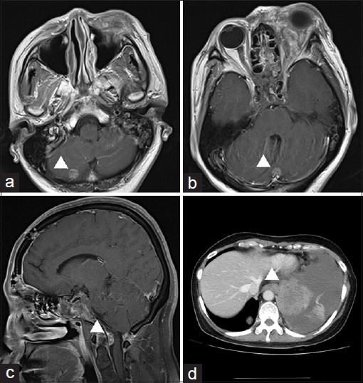
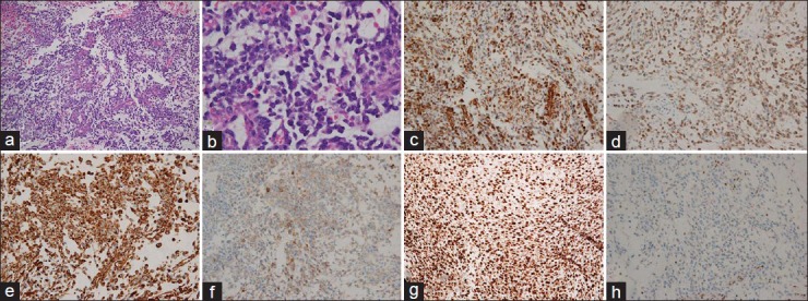
Case description: A 44-year-old female complained of visual disturbance. We performed craniotomies twice and removed partially for each time, but any malignant cells were not found in the specimens. Finally, we determined histological diagnosis from the extended lesion. She died of respiratory failure 17 months after the initial treatment.
Conclusion: AT/RT should be considered in the differential diagnosis of a sellar lesion in adult patients. However AT/RT is rare in adults, the appropriate immunohistochemical evaluation should be performed to diagnose this rare entity.
Keywords: Adult; atypical teratoid/rhabdoid tumor; sellar turcica.
Figures
References
-
- Arita K, Sugiyama K, Sano T, Oka H. Atypical teratoid/rhabdoid tumour in sella turcica in an adult. Acta Neurochir (Wien) 2008;150:491–6. - PubMed
-
- Arrazola J, Pedrosa I, Mendez R, Saldana C, Scheithauer BW, Martinez A. Primary malignant rhabdoid tumour of the brain in an adult. Neuroradiology. 2000;42:363–7. - PubMed
-
- Ashraf R, Bentley RC, Awan AN, McLendon RE, Ragozzino MW. Implantation metastasis of primary malignant rhabdoid tumor of the brain in an adult (one case report) Med Pediatr Oncol. 1997;28:223–7. - PubMed
-
- Balaton AJ, Vaury P, Videgrain M. Paravertebral malignant rhabdoid tumor in an adult. A case report with immunocytochemical study. Pathol Res Pract. 1987;182:713–8. - PubMed
Publication types
LinkOut - more resources
Full Text Sources
Other Literature Sources
