

Regulation of high-voltage-activated Ca2+ channel function, trafficking, and membrane stability by auxiliary subunits
- PMID: 24949251
- PMCID: PMC4059758
- DOI: 10.1002/wmts.93
Regulation of high-voltage-activated Ca2+ channel function, trafficking, and membrane stability by auxiliary subunits
Abstract
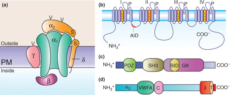
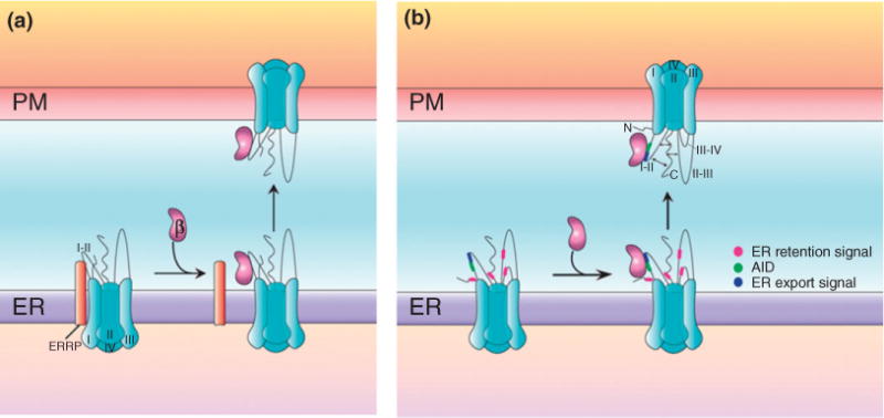
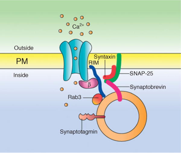
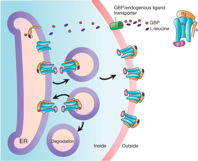
Voltage-gated Ca2+ (CaV) channels mediate Ca2+ ions influx into cells in response to depolarization of the plasma membrane. They are responsible for initiation of excitation-contraction and excitation-secretion coupling, and the Ca2+ that enters cells through this pathway is also important in the regulation of protein phosphorylation, gene transcription, and many other intracellular events. Initial electrophysiological studies divided CaV channels into low-voltage-activated (LVA) and high-voltage-activated (HVA) channels. The HVA CaV channels were further subdivided into L, N, P/Q, and R-types which are oligomeric protein complexes composed of an ion-conducting CaVα1 subunit and auxiliary CaVα2δ, CaVβ, and CaVγ subunits. The functional consequences of the auxiliary subunits include altered functional and pharmacological properties of the channels as well as increased current densities. The latter observation suggests an important role of the auxiliary subunits in membrane trafficking of the CaVα1 subunit. This includes the mechanisms by which CaV channels are targeted to the plasma membrane and to appropriate regions within a given cell. Likewise, the auxiliary subunits seem to participate in the mechanisms that remove CaV channels from the plasma membrane for recycling and/or degradation. Diverse studies have provided important clues to the molecular mechanisms involved in the regulation of CaV channels by the auxiliary subunits, and the roles that these proteins could possibly play in channel targeting and membrane Stabilization.
Conflict of interest statement
Conflict of interest: The authors have declared no conflicts of interest for this article.
Figures
Similar articles
-
An ancestral MAGUK protein supports the modulation of mammalian voltage-gated Ca2+ channels through a conserved CaVβ-like interface.Biochim Biophys Acta Biomembr. 2020 Nov 1;1862(11):183439. doi: 10.1016/j.bbamem.2020.183439. Epub 2020 Aug 16. Biochim Biophys Acta Biomembr. 2020. PMID: 32814116
-
Constitutive activity of the Ghrelin receptor reduces surface expression of voltage-gated Ca2+ channels in a CaVβ-dependent manner.J Cell Sci. 2017 Nov 15;130(22):3907-3917. doi: 10.1242/jcs.207886. Epub 2017 Oct 16. J Cell Sci. 2017. PMID: 29038230 Free PMC article.
-
Transcription Factor Sp1 Regulates the Expression of Calcium Channel α2δ-1 Subunit in Neuropathic Pain.Neuroscience. 2019 Aug 1;412:207-215. doi: 10.1016/j.neuroscience.2019.06.011. Epub 2019 Jun 18. Neuroscience. 2019. PMID: 31220545
-
New Insights in CaVβ Subunits: Role in the Regulation of Gene Expression and Cellular Homeostasis.Front Cell Dev Biol. 2022 Apr 6;10:880441. doi: 10.3389/fcell.2022.880441. eCollection 2022. Front Cell Dev Biol. 2022. PMID: 35465309 Free PMC article. Review.
-
Voltage-dependent calcium channels.Gen Physiol Biophys. 2005 Jun;24 Suppl 1:1-78. Gen Physiol Biophys. 2005. PMID: 16096350 Review.
Cited by
-
Impact of gabapentin on neuronal high voltage-activated Ca2+ channel properties of injured-side axotomized and adjacent uninjured dorsal root ganglions in a rat model of spinal nerve ligation.Exp Ther Med. 2017 Mar;13(3):851-860. doi: 10.3892/etm.2017.4071. Epub 2017 Jan 20. Exp Ther Med. 2017. PMID: 28450909 Free PMC article.
-
Involvement of Parkin in the ubiquitin proteasome system-mediated degradation of N-type voltage-gated Ca2+ channels.PLoS One. 2017 Sep 28;12(9):e0185289. doi: 10.1371/journal.pone.0185289. eCollection 2017. PLoS One. 2017. PMID: 28957379 Free PMC article.
-
Therapeutic Potential of Targeting Regulated Intramembrane Proteolysis Mechanisms of Voltage-Gated Ion Channel Subunits and Cell Adhesion Molecules.Pharmacol Rev. 2022 Oct;74(4):1028-1048. doi: 10.1124/pharmrev.121.000340. Pharmacol Rev. 2022. PMID: 36113879 Free PMC article. Review.
-
Betulinic acid analogs inhibit N- and T-type voltage-gated calcium channels to attenuate nerve-injury associated neuropathic and formalin models of pain.Neurobiol Pain. 2023 Jan 14;13:100116. doi: 10.1016/j.ynpai.2023.100116. eCollection 2023 Jan-Jul. Neurobiol Pain. 2023. PMID: 36687466 Free PMC article.
-
Regulatory Action of Calcium and Calcium Channels in Pain Pathways.Int J Biol Sci. 2025 May 31;21(8):3726-3739. doi: 10.7150/ijbs.110504. eCollection 2025. Int J Biol Sci. 2025. PMID: 40520015 Free PMC article. Review.
References
-
- Perez-Reyes E. Molecular physiology of low-voltage-activated t-type calcium channels. Physiol Rev. 2003;83:117–161. - PubMed
-
- Arikkath J, Campbell KP. Auxiliary subunits: essential components of the voltage-gated calcium channel complex. Curr Opin Neurobiol. 2003;13:298–307. - PubMed
-
- Bourinet E, Soong TW, Sutton K, Slaymaker S, Mathews E, Monteil A, Zamponi GW, Nargeot J, Snutch TP. Splicing of α1A subunit gene generates phenotypic variants of P- and Q-type calcium channels. Nat Neurosci. 1999;2:407–415. - PubMed
-
- Richards KS, Swensen AM, Lipscombe D, Bommert K. Novel CaV2.1 clone replicates many properties of Purkinje cell CaV2.1 current. Eur J Neurosci. 2007;26:2950–2961. - PubMed
Grants and funding
LinkOut - more resources
Full Text Sources
Other Literature Sources
Miscellaneous